
Commentary|Podcasts|April 2, 2026
NeuroOp Guru: Predicting which patients regain vision after optic nerve sheath meningioma treatment
Author(s)Andrew G. Lee, MD, Andrew Carey, MD
Fact checked by: Sheryl Stevenson

Andrew G. Lee, MD, and Drew Carey, MD, review how clinical and imaging findings relate to vision recovery after radiotherapy for optic nerve sheath meningioma.
Advertisement


In this episode of the
Lee is the chair of the Blanton Eye Institute at Houston Methodist Hospital in Texas and a professor of ophthalmology, neurology, and neurosurgery at the Weill Cornell Medical College. Carey is the Neil R. Miller Rising Professor of Ophthalmology in the division of neuro-ophthalmology with the
Carey introduced the clinical challenge: patients with optic nerve sheath meningioma often present with slowly progressive vision loss, and treatment decisions frequently center on whether to observe or proceed with radiotherapy. Because biopsy and surgical removal carry significant risk to the optic nerve, diagnosis is typically based on imaging findings, including MRI features and, in some cases, CT evidence of calcification.
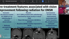
Better baseline vision predicts stronger visual recovery
The discussion focused on a recent single-center retrospective study evaluating visual outcomes after fractionated radiotherapy in patients with presumed optic nerve sheath meningioma.1 Over a 10-year period, investigators identified 26 patients, with 17 ultimately receiving treatment. Carey noted that 59% of treated patients experienced improvement in either visual acuity or visual field outcomes following radiotherapy.
Baseline vision emerged as a key predictor of recovery. Patients who began treatment with better visual acuity—particularly 20/60 or better—were substantially more likely to improve, with many recovering to 20/20 vision. Even among patients without marked central acuity gains, meaningful improvement in visual field defects was observed, including improvement in dense scotomas and mean deviation scores.
Additional factors associated with better outcomes included smaller tumor size and more recent progression, particularly when MRI demonstrated interval shrinkage after treatment. Carey emphasized the practical implication for clinic counseling: delaying treatment until vision has significantly deteriorated may reduce the likelihood of meaningful visual recovery.
The conversation also addressed how these data may help patients who are hesitant about radiotherapy weigh the risks and benefits of intervention. For patients with longstanding severe vision loss, the findings support more tempered expectations for recovery, while reinforcing the importance of earlier treatment in patients with progressive but still salvageable vision.
The discussion reinforces that earlier intervention and stronger baseline visual function are important predictors of visual improvement after radiotherapy for optic nerve sheath meningioma, providing useful guidance for patient counseling and treatment timing in neuro-ophthalmic practice.
Reference
Tien MC, McDonald HM, Wei E, Micieli JA, Margolin EA. Optic nerve sheath meningiomas: a retrospective cohort study comparing outcomes in treated versus observed patients. J Neuroophthalmol. 2025;45(4):466-472. doi:10.1097/WNO.0000000000002309
Advertisement
Related Content
Advertisement


Latest CME


Advertisement
Advertisement