


Imaging
Latest News
Advertisement
Video Series


CME Content


Advertisement

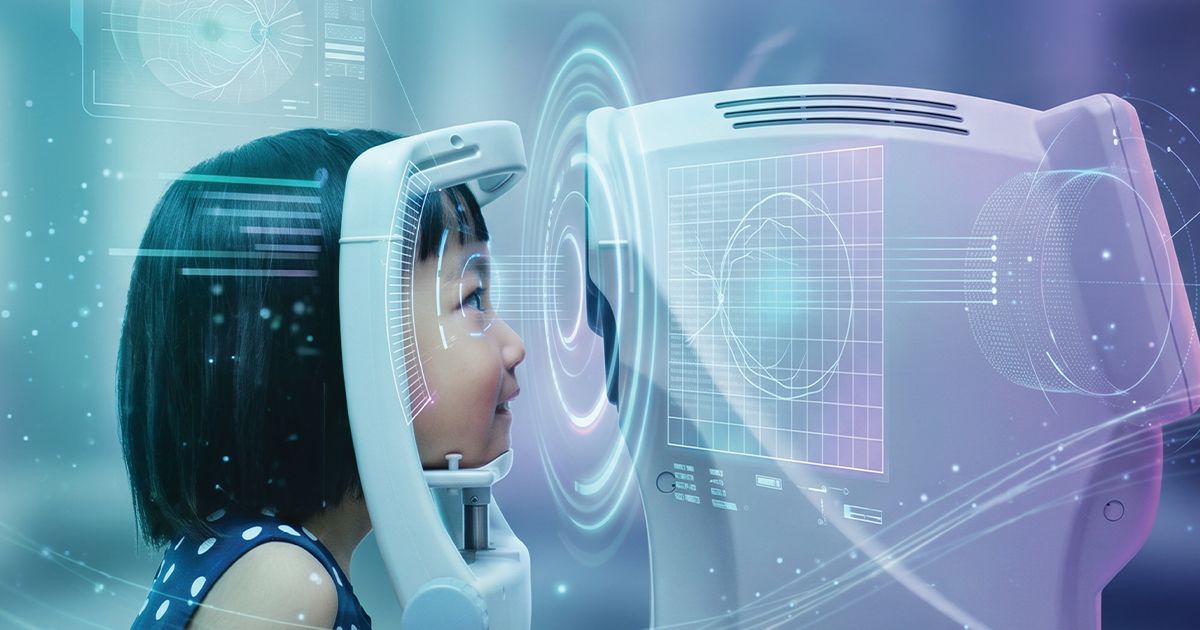
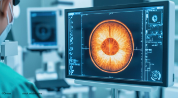
Aixa Alarcon, PhD, and colleagues reported natural image analysis may better predict real-world visual performance of premium intraocular lenses than traditional single-frequency MTF testing, while also capturing the impact of dysphotopsias and residual astigmatism.

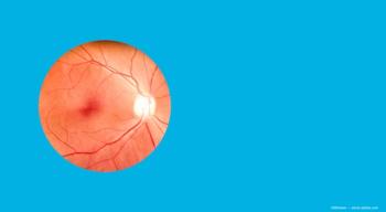
Multimodal imaging, OCT biomarkers, and the case for early diagnosis as treatment options emerge for macular telangiectasia.

At ARVO 2026, Michael Ip, MD, presented findings from a large-scale analysis of OCT biomarkers in patients with diabetic macular edema (DME), leveraging the robust DRCR Retina Network Protocol T dataset.

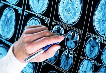
Images showed that Parkinson’s disease is associated with a thicker photoreceptor nuclear layer and a thinner layer of photoreceptor processes.

About 90% of patients are female with obesity and potential hormonal drivers
AI-powered polarized-light retinal imaging separates Alzheimer’s from ALS/FTLD deposits with 96% accuracy, promising low-cost early diagnosis.

Steffen Schmitz-Valckenberg, MD, discusses how optical coherence tomography-based models may enable rapid, noninvasive assessment of functional loss in GA at Angiogenesis 2026.
The 22nd International Scientific Symposium on Ophthalmic Imaging will take place September 18 to 19, 2026 in Singapore, featuring sessions on multimodal imaging, OCT, AI, and next-generation imaging technologies.

China approves ZEISS ARTEVO 750 and 850 ophthalmic microscopes, expanding 3D digital visualization and iOCT-ready workflows for cataract and retinal surgery.

Andrew G. Lee, MD, and Drew Carey, MD, discuss how optic disc cupping after optic neuritis reflects nerve and ganglion cell thinning, not disease type, helping distinguish it from glaucoma.
AI reads infant retinal scans to flag lung disease early, while transient vision loss warns of looming stroke and heart events.

Optoretinography is an emerging technology used to test light-evoked photoreceptor activity.
From artificial intelligence to home monitoring, Joel Schuman, MD, of Wills Eye Hospital, explores the innovations that could change how clinicians detect and treat glaucoma in the new year.

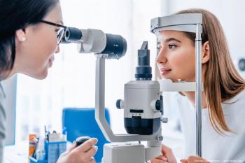
Combining imaging and patient symptoms improves assessment of disease progression.

Dr. Melissa Yuan discusses the impact of weight and postmenstrual age on foveal maturation in preterm infants, highlighting critical insights for neonatal care.
AAO 2025 revealed that true-color widefield imaging, AI-powered home OCT, and refined FAERS analyses are collectively transforming retinal diagnostics into a more precise, continuous, and safety-aware system.


Anat Loewenstein, MD, discusses the transformative impact of home OCT and AI on monitoring retinal diseases at AAO 2025.

Surgeons reflect on milestones that have redefined patient care—and share a glimpse of the advances that promise to shape the next era of eye health.
Deborah A. Ferrington, PhD, highlights how breakthroughs in imaging, AI, and stem cell research are reshaping ophthalmology.

Topcon Healthcare enhances its AI capabilities by acquiring Toku, Inc, aiming to revolutionize eye care with advanced predictive health insights.

The AAO 2025 meeting offered a platform for companies to showcase their latest technologies to advance ophthalmic patient care.
As ophthalmic technologies move at supersonic speed, AI and gene therapy take center stage.
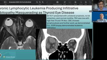
Andrew G. Lee, MD, and Drew Carey, MD, highlight how chronic lymphocytic leukemia can mimic Graves’ orbitopathy, underscoring the importance of a thorough evaluation.
Advertisement
Advertisement