
Blog|Videos|October 22, 2023
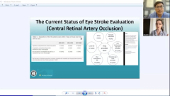
NeuroOp Guru: Two cases of CRAO occurring immediately postoperatively
Author(s)Andrew G. Lee, MD, Andrew Carey, MD
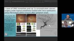
Andrew Lee, MD, and Andrew Carey, MD, sit down on another episode of the NeuroOp Guru to discuss 2 cases of CRAO immediate post-op uncomplicated cataract surgery under topical anesthesia treated with intra-ophthalmic artery fibrinolysis within 6 hours after symptom onset.
Advertisement


Video Transcript
Editor's note - This transcript has been edited for clarity.
Andy Lee, MD:
Hello and welcome to another edition of the NeuroOp Guru. I'm here with Dr. Drew Carey from Johns Hopkins. Hi, Drew.
Drew Carey, MD:
Hey Andy.
Andy Lee, MD:
Today we're going to be talking about central retinal artery occlusion that occurred immediately post-operatively after uncomplicated cataract extraction, under topical anesthesia that was treated with intra-ophthalmic artery fibrinolysis, within 6 hours of symptom onset. Drew, how in the heck did this happen?
Drew Carey, MD:
Yeah, that's a great question, Andy. I just want to be clear for all our listeners out there who don't perform cataract surgery, this is not a common occurrence. So I don't want people freaking out over cataract surgery, this is a super rare event that they just happen to get 2 of these at the same institution. But, cataract surgery happens in older people who are likely to have underlying cerebral vascular risk factors like high blood pressure, diabetes, high cholesterol, maybe they've had a heart attack, maybe they had a prior stroke. And so, you know, they're not different than other people [in] the same age population. But, we tell all patients before surgery, don't take your medicine [and] don't have anything to eat.
So, maybe they get dehydrated and maybe their blood pressure's high, even though for most people, cataract surgery is a pretty painless event. Maybe they had some increased stress and anxiety, and their blood pressure was running extra high. The most common cause of a central retinal artery occlusion is going to be some type of embolic event, usually related to carotid artery stenosis [inaudible] disease. All of those factors together, maybe these 2 patients had an embolic event that got lodged in their central retinal artery occlusion after surgery or immediately during surgery.
Thankfully, they presented very quickly. They must have had an eye doctor or their cataract surgeon told them afterwards. If you ever lose vision, you have to see your eye doctor right away, because that's an eye stroke and could put you at risk for a brain stroke. So, that's kind of what they thought happened. They were very attentive, and I guess they don't patch their patients post op, because otherwise you wouldn't know if you had vision loss. They came back to the eye doctor right away, like super, super fast because the first case they were able to get TPA within less than 3 hours in the second patient less than 6 hours.
That involves recognizing the vision loss, going to see the eye doctor, [and] having a vision exam. They were probably still dilated after cataract surgery, so at least they didn't have to wait for dilating drops to set in. They got OCTs, 1 patient got a fluorescein angiography. They also would have needed a CT of the head to make sure they didn't have any intracranial bleeding before they were treated with TPA. So, all of that got done very, very quickly.
Andy Lee, MD:
These are intra ophthalmic artery deliveries of TPA?
Drew Carey, MD:
Yes. So they were ... they got a cerebral angiogram done. They use itty-bitty, tiny catheters to go up through the carotid artery, and then to make that little turn into the ophthalmic artery. The reason that they do that is it will reduce the risk of TPA spreading into intracranial arteries, and therefore reduce the risk of of having intracranial hemorrhage.
That is the big thing we worry about in anybody who gets TPA, whether it's intravenous, or intra-arterial, is intracranial hemorrhage, because that could be life threatening, as well as other systemic hemorrhages. You can do [a] lower dose of the medication when it's given at the desired location and also help to reduce that spreading to other tissues and reduce the risk of complication. That technique is 1 of the things that's been pioneered, actually by our pediatric oncology fellows for treating retinal blastoma.
They do intra-arterial chemotherapy in the intra-ophthalmic artery. It's a newer technique. It's obviously much more difficult in an infant or toddler than it is doing in an adult. But, our interventional colleagues do have practice with that and thankfully the instruments don't have to be quite as small and adults as they are in children
Andy Lee, MD:
We're pretty convinced these were CREOs not paracentral acute middle maculopathy. Is that right?
Drew Carey, MD:
That is a really great question. Certainly paracentral acute middle maculopathy causes is due to ischemia more at the capillary layer rather than at the arterial layer. You would not expect patients with with PAM to have changes visible on examination. And the other thing that's interesting on the OCT that you can see here in Panel A, there's this outer retinal changes, it almost looks like extension of the photoreceptor layers and maybe trace subretinal fluid.
We don't actually see whitening of the, or hyper reflectivity of the of the inter retinal layers, probably because we got to it before it reached infarct level. I think the vision loss to count fingers would be really atypical for PAM. And, and the diffuse vision loss that that's described would also be a typical for PAM. PAM usually is going to be more of a paracentral scotoma. And visual acuity often very good unless it involves Papillomacular Bundle.
Andy Lee, MD:
And what do you think about this timing? Five and a half hours versus 4 and a half hours? You have a time that's different than regular stroke patients or is it variable?
Drew Carey, MD:
Well, we don't know what the time cutoff is for treating central retinal artery occlusions. There are a couple prospective trials going on in Europe that will hopefully give us more information. I think if you ask, you know, a stroke specialists, TPA, intravenous TPA, was originally 3 hour time window. And then they've expanded that in patients, they felt were low risk to 4 and a half and sometimes 6 hours.
We know for intra-arterial TPA, that is a longer time window than intravenous there, those patients are at lower risk of hemorrhagic conversion, which is really the cutoff for that time window. And because you're giving it again, kind of focally, as opposed to the whole brain, and in these patients, you know, they seem to be probably lower risk, because again, we're not perfusing the whole brain with TPA and not giving them that full risk of intracranial hemorrhage.
I think if you asked a stroke specialist, you know, how long do you have to reverse ischemic damage to a brain that has a true, you know, a true ischemic deficit. I think most people were telling you, you know, if it's infected, that part of the brain, you're probably not recovering, what we're really trying to recover the ischemic area around it, so that patients can get as much recovery as possible. And I think, you know, in the retina, that's, that's kind of a similar thought.
And most people would have assumed that maybe you have have 20 to 30 minutes to try and recover retinal function. If it's a true infarct. I think there's probably degrees to central retinal artery occlusion, you know, if there's enough blood flow to disrupt neurologic function in the retina, it's ischemic. And if that persists long enough, you're not going to recover anything.
But if it's not a full infarct, then you have time for recovering and we don't really know what that is, we know from the old studies out of the University of Iowa, in monkeys, where they would clamp the central retinal artery surgically, you know, behind the eye. And using ERG studies, the meantime for full recovery of ERG function was about 90 minutes. Now, does that mean they're gonna have normal vision? No, there's probably some, you know, permanent retinal infarcts in there, but not the whole retina.
And you have to kill off at least 25% of the retina to see a change on a on a full field ERG. I think you know, when you have a patient like this, who's who's essentially blind in the eye, you're happy to get any vision back these patients recovered to 20/20 and 20/25. And I'm, you know, I'm thrilled to see that there's that kind of recovery that can happen at 3 hours and almost 6 hours after onset of vision loss.
You know, there may be some patients where you don't get that level of recovery back because maybe it's a full occlusion and then you're gonna see earlier stages of infarct but I think getting you know, anything, you know, that's that's reading level of vision, most people would consider that pretty normal vision. We don't have the fields. You know, we don't we don't have microperimetry or multifocal ERG, so we don't know if it's 100% normal, but that's really good, you know, functional vision.
And I'm ecstatic even, you know, it's only 2 cases and you said, "Oh, Dr. Carey, you know, some patients recover vision anyways, if you don't do anything," you know, if it's me get, give me the TPA. So I think, you know, the big challenge going forward is how do you get the patients in the door in time to do this? And, you know, and then reduce that door to needle time?
Andy Lee, MD:
What are you guys doing at Hopkins in terms of intravenous versus intraophthalmic?
Drew Carey, MD:
I don't think we've had anybody come in the door, in what you would consider the TPA window for central retinal artery occlusion. For brain strokes. You know, usually they'll do intravenous TPA, if you get them within that 3 to 4 and a half hour window, depending on, do they have diabetes are there other risk factors. You know, for patients who have significant persistent deficit, and they're beyond that they're in that 6 to 12 hour window. Most of the time, you know, the stroke team will consider intra-arterial TPA or intravascular thrombectomy for patients with brain strokes.
Andy Lee, MD:
Did they say what they thought the mechanism was for topical anesthesia. Is it IOP or bad luck? Or what was the cause here?
Drew Carey, MD:
Oh, I don't think that topical anesthesia is felt to contribute to the central retinal artery occlusion. It is to distinguish patients who might have retrobulbar anesthesia, where you could have increased intraorbital pressure that compresses the central retinal artery occlusion, or if the patients were to receive retrobulbar epinephrine as part of the anesthesia that can induce vaso spasms.
Or if you have a sickle cell patient it can induce sickling. And that could be those mechanisms could contribute to central retinal artery occlusion. So that noting that it was topical anesthesia was to take away other mechanism that could have resulted from some type of retrobulbar mechanism or injury in a traumatic injury from a retrobulbar needle or medication.
Andy Lee, MD:
So Drew, what do you think the take home message is for our audience about this paper?
Drew Carey, MD:
I think the probably the 2 most important take home messages is that if, you know I think education can can work. If we tell our patients if you have sudden painless vision loss and an eye that might be nice stroke and you have to be seen right away.
Patients will listen to you and that's really important if we're going to hope to treat central retinal artery occlusion. So I think as as eye doctors, we can move the the needle, pun intended, on by educating our patients. And the second one is that it appears that intra-arterial treatment with TPA for central retinal artery occlusions, if given properly may restore vision and may be safe. And stay tuned as we get prospective data from randomized trials coming hopefully in the near future. And this may be coming to an institution near you.
Andy Lee, MD:
Well Drew, as always a pleasure chatting with you. That concludes yet another edition of the NeuroOp Guru we'll see you guys next week. Thanks Drew.
Advertisement
Related Content
Advertisement


Latest CME


Advertisement
Advertisement