


Ted Leng, MD, discusses his IRIS Registry analysis presentation regarding variations in vitreoretinal physician utilization of ancillary testing.
Ted Leng, MD, discusses his IRIS Registry analysis presentation regarding variations in vitreoretinal physician utilization of ancillary testing.
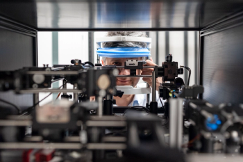
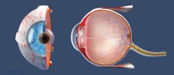
A team of scientists have developed a two-photon excited fluorescence scanning laser ophthalmoscope, an instrument that allows viewing the biochemistry of vision in the living eye in real time.

Approach shows potential as a promising second-line screening tool for patients with diabetes.

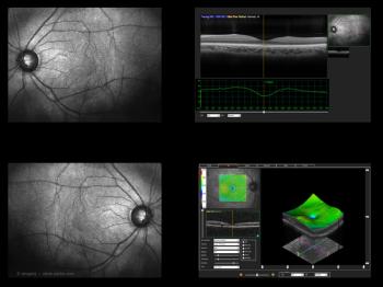
SD-OCT is providing reproducible, high-quality, registered images to assess the treatment response in macular disease.
Widefield fundus autofluorescence detects subtle findings, which can result in better outcomes.
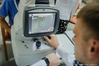
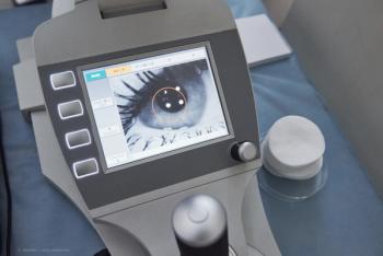
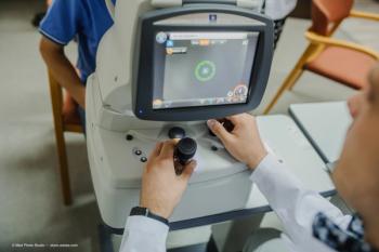
A pilot study tests the technology to obtain remote fundus imaging of patients.

Investigators hoping to develop a novel artificial intelligence model.
Proposal hints at the use of generative adversarial network (GAN) for applications in macular surgery
Diagnostics, imaging, and therapeutics pave the way for better outcomes.
Retinal and choroidal changes were observed in study.
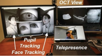
Pilot study tests the technology to obtain remote fundus imaging of patients.
Patients can get real-time disease monitoring with self-operated device.
Adolf Fercher's 2D image of fundus paved way for today’s imaging technique.

Investigators examine usefulness in refractive surgery evaluations.

Anat Loewenstein, MD, MHA, discusses how artificial intelligence can optimize optical coherence tomography.
Scanning laser ophthalmoscopy offers improved view of posterior segment.
Philip Rosenfeld, MD, PhD, presents results from a natural history study of patients that results in recognition of hypertransmission defects on en face OCT.
Tool for quantifying measures may predict outcomes for patients.
Option detects treatment-naive nonexudative macular neovascularization in eyes with dry age-related macular degeneration

Investigators have found that optical coherence tomography findings could prove to be a diagnostic tool for COVID-19.
First step is ruling out ocular nystagmus, saccadic disorder.
Dual imaging may reduce costs, unnecessary referrals, telehealth study results show.
Measures of inflammation can predict treatment outcomes for patients.

Retinal imaging tests are providing material to train and test decision-support systems.