


Imaging
Latest News
Advertisement
Latest Videos


CME Content


Advertisement
More News
In celebration of Ophthalmology Times 50th anniversary, we asked leading experts in the field what they see as the biggest innovation in ophthalmology in the last 5 decades.
The company announced it has successfully started imaging patients with its first prototype of its device using both near-infrared and green modes.
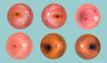
Ophthalmologist offers insights into the value of technology in diabetic retinopathy.
The UK-based company will debut the tool, called Dr.Noon CVD, at two conferences in March.
The device recently received 510(k) clearance from the US Food and Drug Administration.

The Sentinel Camera aims to address critical gaps in retinal disease screening by offering a portable device that captures high-quality images that require no dilation of the eye.

The AI-READI project aims to establish fair, equitable, and ethical access to big data, enhancing artificial intelligence’s ability to diagnose systemic diseases and drive progress in ophthalmology research.

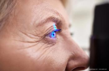
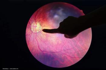
A look at the latest advances from flash photography to AI-driven OCT

With cases of syphilitic uveitis on the rise, recognizing its varied presentations—including optic disc edema, which may appear without other significant eye inflammation—is increasingly important for timely diagnosis and treatment.

Hyaluronic acid fillers are used for undereye volume correction, detailing aging effects on facial anatomy, common complications, and optimal injection techniques. Imaging tools such as MRI can prove to be beneficial for patient assessment.

Perimeter Medical Imaging AI has been leveraging the technology to improve breast cancer surgeries and reduce the number of patients requiring repeat surgery.

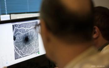
In a study, a team of Korean researchers developed an AI model using OCT images to predict neovascular AMD treatment outcomes after anti-VEGF injections. The model highlights AI’s potential in personalized ophthalmic care.

A study by a team of Chinese researchers highlights the potential of optical coherence tomography angiography (OCTA) in detecting retinal microvascular damage in renal hypertension patients. OCTA identifies reduced vascular density in key retinal regions, offering a noninvasive tool for early diagnosis and systemic disease management.

News
A recent cohort study revealed low adoption of FDA-approved AI-based diabetic retinopathy detection, with less than 5% of diabetic patients receiving ophthalmic imaging. Researchers emphasize the need for improved awareness, cost-effectiveness, and integration to increase diabetic retinopathy screening rates.
News
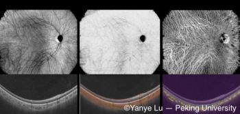
Peking University researchers have developed a deep learning-based, noninvasive choroidal angiography method that enables detailed 3D visualization of choroidal vessels from OCT scans. This technique could improve diagnostics for retinal diseases like macular degeneration, offering a safer alternative to traditional methods.

A home self-imaging device can provide ophthalmologists with actionable insight between office visits for AMD

Researchers find a strong link between Alzheimer's disease and retinal thinning

News
The award from the National Institutes of Health will enable a team of researchers to investigate Alzheimer and Parkinson progression through the eye.

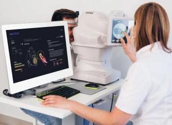
CVD could impact physicians' ability to identify choroidal melanoma from choroidal nevus.

The nOCT allows for detailed visualization of the brain's vascular anatomy.
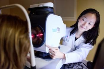
TFI employs spectral interference technology to image and map the corneal surface.

The companies are teaming up to reduce care gaps and improve eye and vision health by bringing diabetic retinopathy and other eye disease screening services to patients in their own homes and workplaces.

Technology offers increased usability, safety, and accuracy in MIGS procedures.

Comanagement is a key component for treatment of disease.
Advertisement
Advertisement
Trending on Ophthalmology Times - Clinical Insights for Eye Specialists
1
Eyes on May 2026: A month of durable therapies, a regulatory reversal, and rethought echanisms
2
Reasoning prompts sharpen multimodal AI on bilingual ophthalmology exam questions
3
TECNIS PureSee IOL launches in US following FDA approval for cataract surgery
4
Beyond the Walls: Leading with love, loyalty, and a team first philosophy with Vance Thompson, MD
5