


The therapy may offer novel treatment strategy with unique mechanism, modality for patients diagnosed with dry age-related macular degeneration.
The therapy may offer novel treatment strategy with unique mechanism, modality for patients diagnosed with dry age-related macular degeneration.
On this week's episode of the NeuroOp Guru, Andy Lee, MD, and Elizabeth Fortin, MD, compare the 1.5 Tesla magnet and the 3.0 Tesla magnet for MRI.
The advanced swept-source OCT technology allows visualization of the variations in the normal changes and those associated with aging in the posterior vitreous.

When paired with SD-OCT, the device also can be used to evaluate photoreceptors.

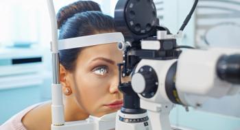
According to researchers, functional assessments and education are critical to an early diagnosis.

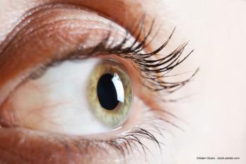
According to a new study from New York Eye and Ear Infirmary of Mount Sinai, a new, non-invasive ocular imaging method may be able to detect an early indicator of glaucoma in time to prevent disease progression and vision loss.
Flavoprotein fluorescence could serve as a new biomarker, according to a Mount Sinai study.

Gareth Lema, MD, PhD, discusses an innovative eye stroke program at Mount Sinai Health System that involves collaboration among ophthalmologists, the stroke service, and emergency department physicians.
According to the company, new imaging options presenting opportunity for better outcomes for patients.
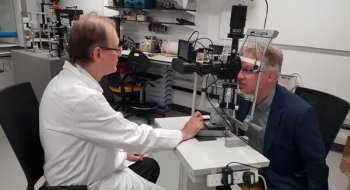
Researchers at the University of Strathclyde have developed an affordable device which takes 3D images and could shift the landscape of eye screening and treatment around the world.

The study investigates 3-year intravitreal implant in eyes with active noninfectious posterior uveitis.
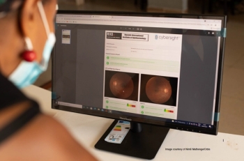
The research from eye care nonprofit Orbis International focuses on diabetic retinopathy in Rwanda, where associated vision complications from diabetes are growing.
With clearer imaging and enhanced resolution, the new OCT approach could improve medical diagnostic imaging.
The rare disorder is often a delayed or misdiagnosed condition, resulting in unnecessary referrals and imaging.
A new technique will allow fast and non-invasive assessment of the physiological state of the retina. This could be a real breakthrough in the treatment of eye diseases.

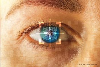
In a patient survey, 66% of respondents said AI plays a large role in their diagnosis and treatment and thought it was important.

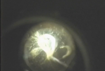
TAKE-HOME: This affordable device allows the view through a slit lamp to be shared in real time or recorded, for teaching and reference purposes.

A team of investigators from Pohang University of Science and Technology has found that conjunctival goblet cell examination is important for the precise diagnosis and effective treatment of ocular surface diseases; however, CGC examination has not been possible until now due to lack of non-invasive devices.
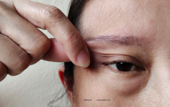
Assessing cornea status can aid early recognition of disease-related changes

Researchers from the Department of Ophthalmology at the University Hospital Bonn and Deutsches Zentrum für Neurodegenerative Erkrankungen suggest that assessments of the eye’s retina could help to detect a loss of brain substance, i. e. “brain atrophy.” The findings are based on data from the Rhineland Study.

Mary Durbin, PhD, Chief Scientific Officer at Heru, discusses the benefits and capabilities of the various testing modalities available in 1 wearable platform.

During her talk at Angiogenesis, Dr. Loewenstein outlines how artificial intelligence could revolutionize diabetic retinopathy screening.
Robert L. Stamper, MD, speak with Ophthalmology Times®' David Hutton to discuss his presentation at the Glaucoma 360 event in San Francisco, where he presented an update on OCT-Angiography and its role in detecting the density of the capillaries in the macula.
The GMOPC was established in February 2020 at an inaugural meeting in Los Angeles, California, as an investigator-initiated clinical research study, with Heidelberg Engineering as an industry partner.