
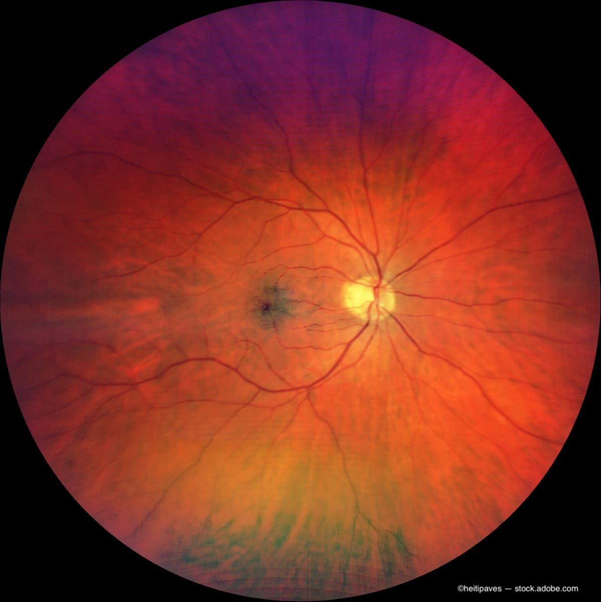











Researchers at Mass Eye and Ear have demonstrated that retinal imaging can help predict a person’s risk of developing ocular, neuropsychiatric, cardiac, metabolic, and pulmonary diseases.
Researchers at Mass Eye and Ear have demonstrated that retinal imaging can help predict a person’s risk of developing ocular, neuropsychiatric, cardiac, metabolic, and pulmonary diseases.
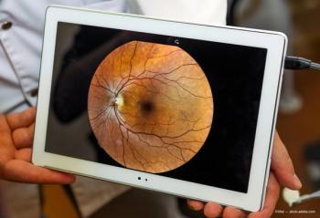
While the potential for fundus images is recognized, the methodology and clinical application must be improved.
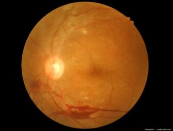
The kiosk is the first to offer self-service retinal imaging, empowering individuals to capture their own images.
Varun Chaudhary, MD, FRCSC, addressed the big questions in treating patients with retinal vascular diseases at the Retina 2024 meeting in Maui.
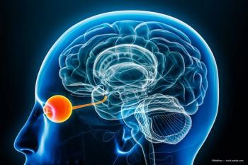
The goal is to understand how individuals with CVI interact with their visual world
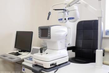
Scientists have developed a versatile imaging system that will help diagnose pathologies in the eye fundus.
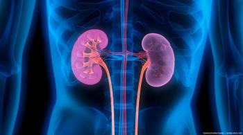
The kidney and eye are structurally and functionally similar, and the diseases of the organs may present similarly and via common pathways.
A Q-switched, 532 nm-wavelength, frequency-doubled Nd:YAG laser, the Eagle is intended for use in performing selective laser trabeculoplasty.

ROP can be a disease that affects both the retinal and choroidal vasculatures.
The company has come into agreement with IQVIA Services Japan G.K. as well as AUROLAB.
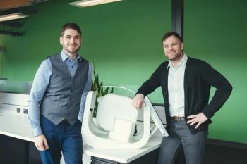
The platform technology can be adapted to assess a vast array of biomarkers present in the eye.

The investigators use mindfulness-based cognitive therapy (MBCT) on patients who have never previously practiced the technique, with the aim of possibly re-training their brains to remove their static-type images.
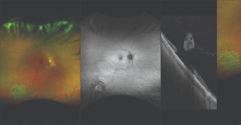
Technology is an option for evaluating, documenting certain types of retinal pathology.

The collaboration, fueled by a $5 million donation from Knights Templar Eye Foundation (KTEF), will introduce a free and open VR simulation program for ophthalmologists and trainees worldwide.

The goal is to evaluate neonates with retinopathy of prematurity (ROP) who met only 1 screening criterion with the goal of easing the burden of ROP screening.
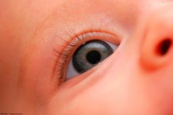
Investigators have proposed a new algorithm, TWO-ROP, which they describe as “a new and simple screening protocol with a two-tiered impatient and outpatient approach.”
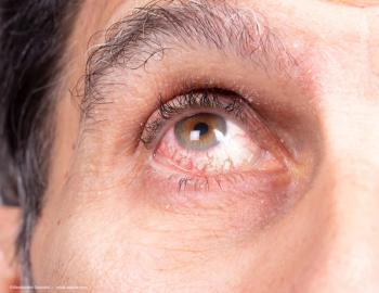
Physician discusses 5 keys to achieve optimal results for patients
The investigators performed clinical examinations and obtained fundus photographs and inferior optical coherence tomography (OCT) images for both treatments at baseline and on days 1, 3, 8, 15, and 56 after the injections.

Researchers at the Warsaw, Poland-based International Center for Translational Eye Research decided to change that by introducing a new imaging method derived from optical coherence tomography. This led to the creation of even more advanced spatio-temporal optical coherence tomography.

The collaboration will drive an effort to develop predictive analytics to enable early detection of eye diseases and more personalized treatments to prevent vision loss for people with serious eye diseases.

Altris also has advanced AI models to enable vendor-neutral retina layers segmentation as well as detection and visualization of more than 70 retina conditions such as dry and wet age-related macular degeneration, geographic atrophy, diabetic retinopathy, diabetic macular edema and central retinal vein occlusion.
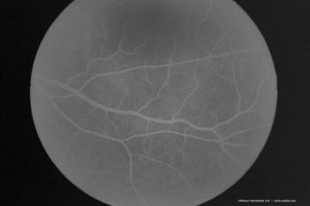
Investigators note a supply shortage led to use of half a dose for imaging purposes.
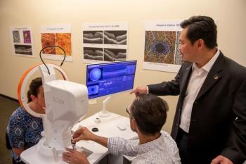
The Lasker-DeBakey Clinical Medical Research Award, dubbed "America’s Nobel," recognizes the wide use of optical coherence tomography to manage eye disease, prevent blindness.

Rather than speculating in a vacuum about the future of remote monitoring in ophthalmology, it is better to explore how other specialties in medicine adopted and integrated remote monitoring into their care plan frameworks.
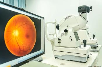
The company has released a video presentation that highlights combining genetic markers with high-resolution retinal imaging to assist in the detection and treatment of blindness and multiple systemic diseases.