


Imaging
Latest News
Advertisement
Latest Videos


CME Content


Advertisement
More News

Recapping the Heidelberg Engineering International SPECTRALIS Symposium—and Beyond.

Advanced imaging and awareness of systemic risk factors are essential.
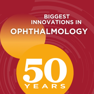
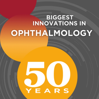
Ophthalmology celebrates 50 years of groundbreaking innovations, highlighting anti-VEGF therapies, OCT imaging, and future advancements in eye care.
Integrating IRIS technologies will allow Topcon to improve retinal disease diagnostics and referrals from primary care providers.

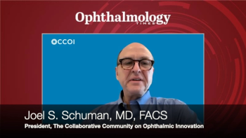
President Joel S. Schuman, MD, highlights the group’s mission and vision for the future—uniting 17 working groups and over 80 stakeholders.
At the 2025 International SPECTRALIS Symposium—And Beyond (ISS), Chauhan discussed how two-photon microscopy enables precise, non-invasive monitoring of retinal ganglion cell function in living subjects.

From Nobel laureates to AI-driven research, the Wilmer Eye Institute honors a century of transforming vision science and care.
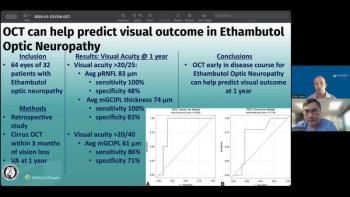
Andrew G. Lee, MD, and Drew Carey, MD, discuss how baseline optical coherence tomography parameters can help ophthalmologists counsel patients and make more informed decisions after ethambutol-associated optic neuropathy.
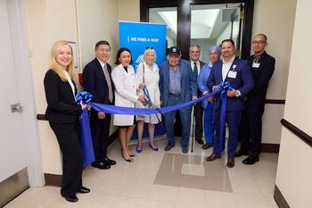
Q&A: Kira Manusis, MD, on how the Center for Refractive Solutions is redefining patient care at NYEE
As its director, Manusis outlines the facility’s role in delivering advanced, personalized solutions to improve vision and patient experience.

ZinserLab will focus on accelerating the development of imaging technologies in eye care.
With detailed imaging and cognitive data, the Northern Ireland Cohort for the Longitudinal Study of Aging highlights the potential of integrating eye scans into broader health research.

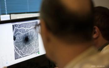
Optical coherence tomography markers—like the ellipsoid zone—are reshaping clinical trials in intermediate age-related macular degeneration, as highlighted at the 2025 International SPECTRALIS Symposium — And Beyond (ISS).

Fluorescence lifetime imaging ophthalmoscopy is emerging as a valuable tool to reveal previously hidden links between retinal changes and systemic disease.
The center aims to optimize patients' vision with a range of cataract and corneal refractive procedures and minimize reliance on glasses and contacts.

Deep learning discerns IIH, NAION, and normal eyes using single fundus image.

With 35+ lectures, a NASA keynote, and a river cruise through historic Heidelberg, this year’s symposium blends science and scenery

Jay Chhablani, MD, explains how 3D choroidal vessel segmentation transforms ophthalmology, enhancing disease diagnosis and treatment through advanced imaging technology.

Compensation techniques in swept-source optical coherence tomography angiography improve accuracy by correcting signal loss from drusen and other artifacts

Researchers introduce a multistage dual-branch network to improve accuracy and efficiency
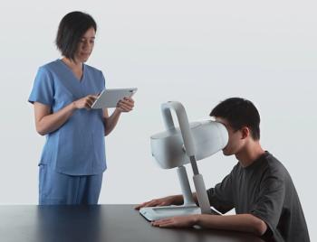
The company announced it has successfully started imaging patients with its first prototype of its device using both near-infrared and green modes.
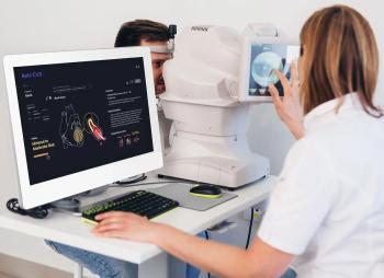
Ophthalmologist offers insights into the value of technology in diabetic retinopathy.
The UK-based company will debut the tool, called Dr.Noon CVD, at two conferences in March.
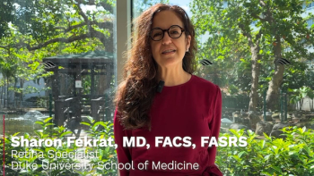
At the Envision Summit 2025 in San Juan, Puerto Rico, Sharon Fekrat, MD, FACS, FASRS talked about multi-modal retinal and choroidal imaging to diagnose and identify neurodegenerative diseases using machine learning.
Advertisement
Advertisement
Trending on Ophthalmology Times - Clinical Insights for Eye Specialists
1
Eyes on May 2026: A month of durable therapies, a regulatory reversal, and rethought echanisms
2
Reasoning prompts sharpen multimodal AI on bilingual ophthalmology exam questions
3
TECNIS PureSee IOL launches in US following FDA approval for cataract surgery
4
Beyond the Walls: Leading with love, loyalty, and a team first philosophy with Vance Thompson, MD
5