


Imaging
Latest News
Advertisement
Latest Videos
CME Content


Advertisement
More News

The mapping method revealed prominent microscopic abnormalities consistent with diabetic retinopathy.

Alternate methods of screening may lead to earlier diagnosis of disease.

Joseph Izatt was a skilled researcher and inventor who played a foundational role in the development of optical coherence tomography.

Contrary to the traditional understanding that this disorder remains stable, long-term structural changes were observed through spectral-domain optical coherence tomography (SD-OCT) imaging.
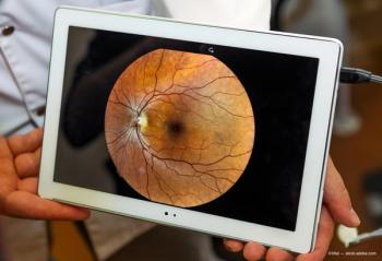
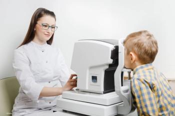
Mobile cameras allow University of Colorado ophthalmology residents working in hospital settings to conduct more comprehensive and efficient patient evaluations.

According to the company, 2,500 clinical and peer-reviewed studies that span 235 disease states have demonstrated the clinical utility of Optos technology.
Multimodal technology bridges the gap between advanced age-related macular degeneration (AMD) biomarkers and clinical features.

Researchers at Mass Eye and Ear have demonstrated that retinal imaging can help predict a person’s risk of developing ocular, neuropsychiatric, cardiac, metabolic, and pulmonary diseases.
While the potential for fundus images is recognized, the methodology and clinical application must be improved.
Varun Chaudhary, MD, FRCSC, addressed the big questions in treating patients with retinal vascular diseases at the Retina 2024 meeting in Maui.

The goal is to understand how individuals with CVI interact with their visual world

A Q-switched, 532 nm-wavelength, frequency-doubled Nd:YAG laser, the Eagle is intended for use in performing selective laser trabeculoplasty.

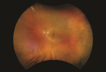
ROP can be a disease that affects both the retinal and choroidal vasculatures.
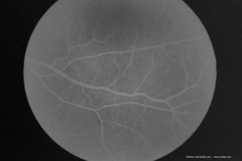
Technology is an option for evaluating, documenting certain types of retinal pathology.

The goal is to evaluate neonates with retinopathy of prematurity (ROP) who met only 1 screening criterion with the goal of easing the burden of ROP screening.

Physician discusses 5 keys to achieve optimal results for patients
Investigators note a supply shortage led to use of half a dose for imaging purposes.

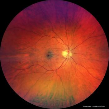
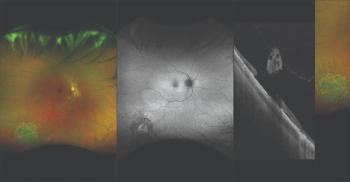
Researchers combine blue reflectance, red/green 200° ultrawidefield images

Oluwatosin U. Smith, MD, shares why she is excited for the conference and why it is relevant for today’s ophthalmologists and optometrists.

At the 2023 ASRS meeting in Seattle, Washington, Aaron Lee, MD, spoke with our team about his research and how changes are making new training possible. He shared how research and new techniques are expanding the options for deep learning in ophthalmology.

Trainees learn to recognize key pathologies seen in fundus examinations, OCT images
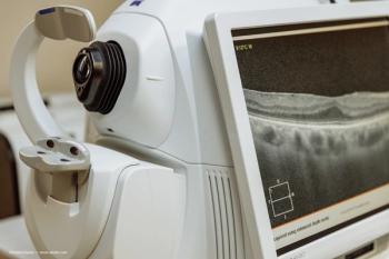
OCT methods are invaluable in the assessment of patients with the condition.
According to company officials,the new image modality provides additional retinal visualization to our customers as they manage their patient’s treatment and ongoing care.
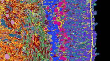
A team of researchers has now developed an approach to gather and compile a great deal of information about organoids and their development. The team applied its approach to the organoids of the human retina, which they derived from stem cells.

Tips to help eye care providers navigate the maze of insurance issues.
Advertisement
Advertisement
Trending on Ophthalmology Times - Clinical Insights for Eye Specialists
1
Tinlarebant NDA submission completed for Stargardt disease type 1, targeting first approved therapy
2
FDA accepts Outlook Therapeutics' resubmitted BLA for ONS-5010 (LYTENAVA)
3
Study finds mixed real and synthetic eye images enhance ocular AI performance
4
Recognizing biomarkers of Alzheimer Disease using OCTA
5