







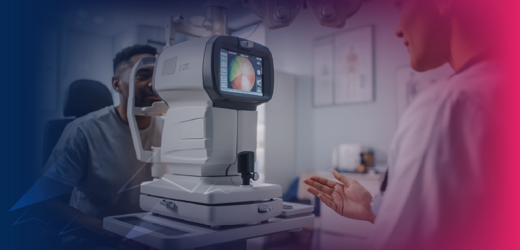
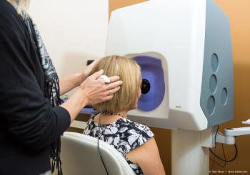
Physicians should evaluate which options best meet the needs of their practices.
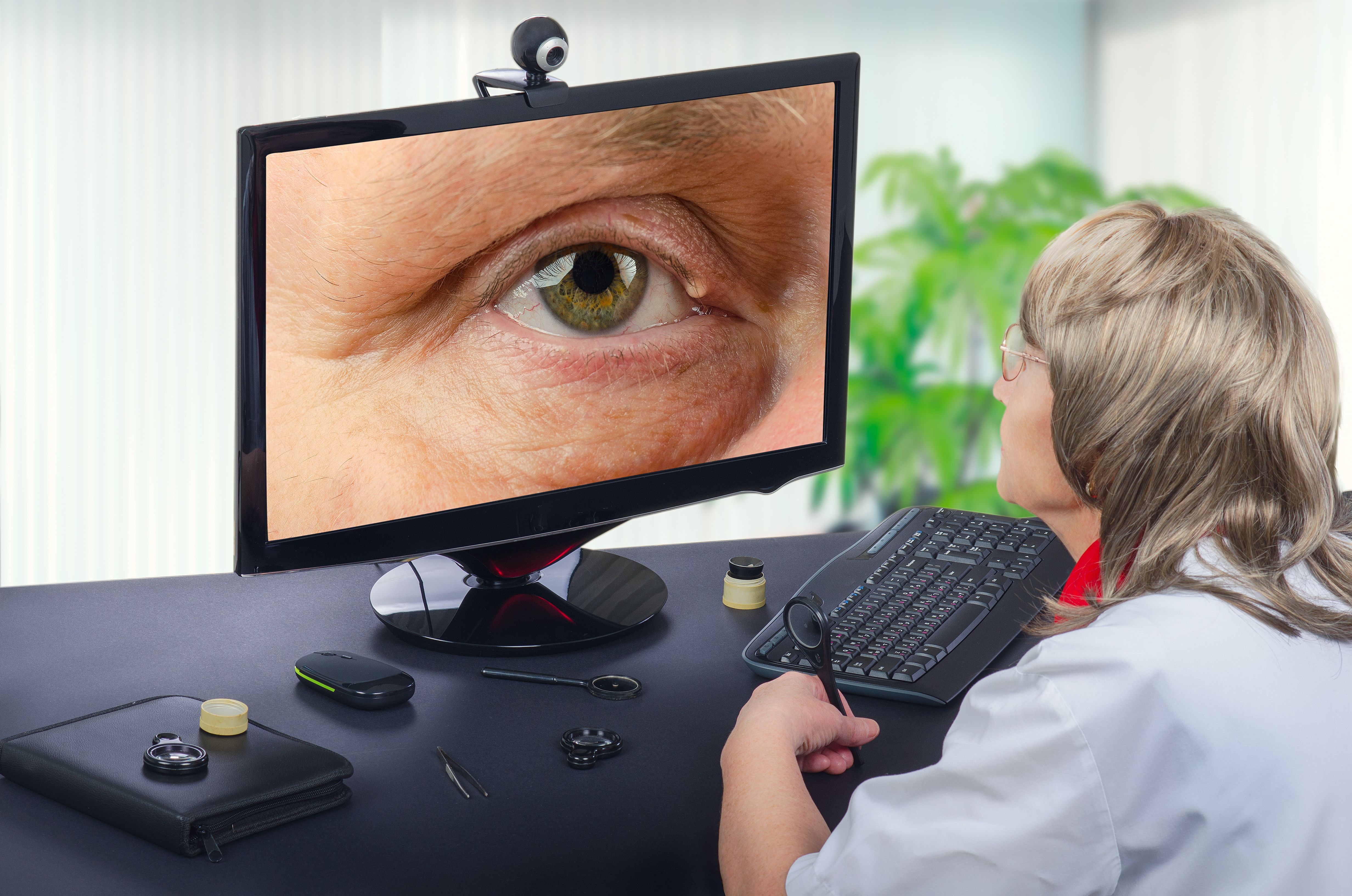
Physicians should evaluate which options best meet the needs of their practices.
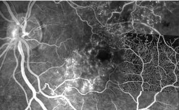
Tool can help physicians detect, monitor ophthalmic disease in their patients.
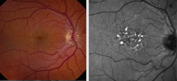
Key is to recognize usual patterns for systemic and topical medications.
Emerging technology may facilitate individualized care, disease management.

Higher levels of 3 macular pigments minimize risk of age-related disease.

Imaging ciliary muscle details mechanism of accommodation, effects of aging .

Process eliminates need for expensive in-person examination of patients without DR.

Study finds imaging-based formulas are less accurate in post-myopic LASIK eyes.

Dilation may be an important step to reduce the rate of ungradable images
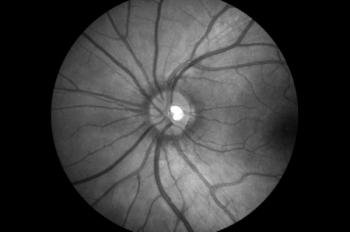
High-quality images can be useful for early diagnosis, prognosis, monitoring
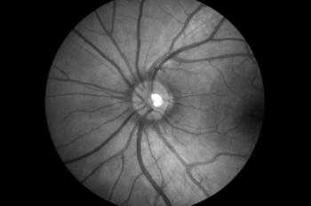
High-quality images can be useful for early diagnosis, prognosis, monitoring
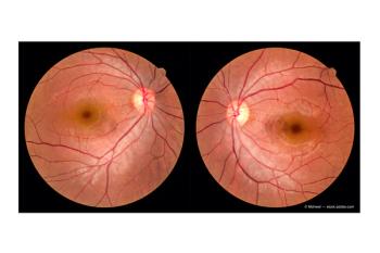
Study designed to identify patterns in abnormalities and differences characteristic of disease.
Results of a prospective trial show the tool is ready for clinical use and potential deployment for remote telemedical use.

Study explores correlations between functional and structural tests
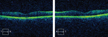
Investigators find value in screening for macular disease in preoperative evaluation
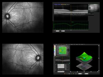
Investigators tested the hypothesis that changes in certain areas of the macular are more relevant to AMD. What did their findings reveal?
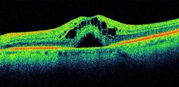
The Audacious Goals Initiative of the National Eye Institute is focused on developing noninvasive imaging advances to help patients with vision loss.
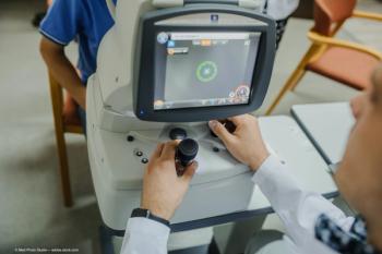
These imaging technologies can help with assessing unexpected structural and vascular retinal and choroidal changes in active and post-treatment toxoplasmic retinochoroiditis.

A study of adult patients with diabetes found that bowtie-shaped polarization artifacts do not serve as reliable clinical biomarkers for center-involving diabetic macular edema. What might this mean for clinicians?

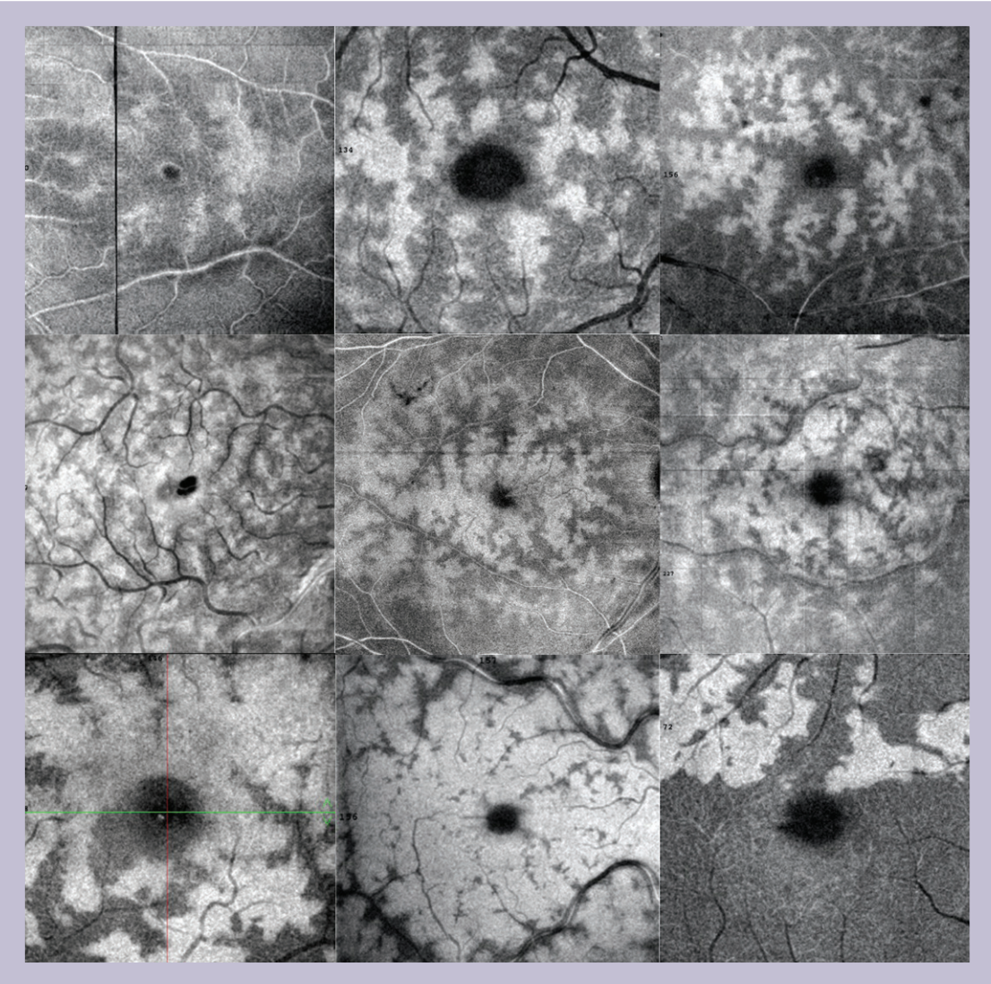
Fundus autofluorescence is a helpful tool for diagnosing central serous choroidopathy and planning treatment.
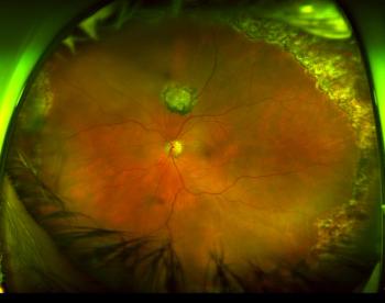
Physicians can discover, diagnose, document, treat diseases that might go undetected
Researchers seeking fluid features, retinal integrity measures to test next step in image analysis and image interrogation in clinical trials.
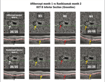
When used one week after injection, optical coherence tomography angiography can help clinicians determine how effective their treatment has been on neovascular membrane reperfusion.