








The retina is a peripheral part of the central nervous system and shares many similarities with cerebral brain matter.

The retina is a peripheral part of the central nervous system and shares many similarities with cerebral brain matter.

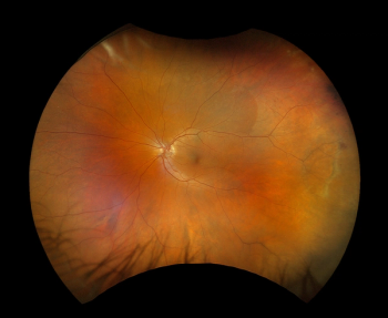
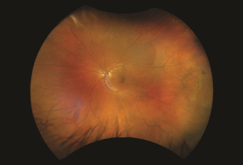
Optos announced the first ultra-widefield color rgb (red/green/blue) image. This new image modality is captured simultaneous to the optomap color rg image.

According to company officials,the new image modality provides additional retinal visualization to our customers as they manage their patient’s treatment and ongoing care.

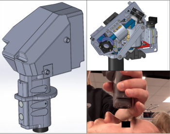
This medical device is the first retinal eye-movement monitor for non-invasive, objective clinical assessments.

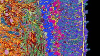
A team of researchers has now developed an approach to gather and compile a great deal of information about organoids and their development. The team applied its approach to the organoids of the human retina, which they derived from stem cells.

Dierck Hillmann, PhD, was interviewed by our onsite team at the 2023 ARVO annual meeting where he shared research on holographic optical coherence tomography (OCT).

At the 2023 ARVO annual meeting in New Orleans, our on-site team caught up with T. Y. Alvin Liu, MD, to discuss predictive AI and its application in ophthalmology and screening of the eyes.

David Szanto, a medical student at Stony Brook, and working with the Department of Neurology at the Icahn School of Medicine at Mount Sinai, describes how an algorithm finds recurring patterns in the visual fields and the ganglion cell-inner plexiform layer thicknesses, and helps clinicians understand how prevalent those patterns are throughout the entire data.

Tips to help eye care providers navigate the maze of insurance issues.

The various aspects of home monitoring for patients with age-related macular degeneration are discussed in this point/counterpoint by Prof. Rufino Silva, University of Coimbra / Coimbra Hospital and University Centre, Portugal, and Univ.-Prof. Dr. med. Robert P. Finger, PhD, Department of Ophthalmology, University of Bonn, Germany, during the 14th annual Congress on Controversies in Ophthalmology in Lisbon, Portugal.

Imaging biomarkers are useful to determine the degree of EZ attenuation in the fovea.

Delegates will analyze the most relevant and controversial issues within the field of ophthalmology at this year’s congress in Lisbon, Portugal, from March 24 to 25.

This subtype of artificial intelligence technology may have long-range implications for clinical trials.

Artificial intelligence (AI) with ultra-widefield fundus autofluorescence images may be helpful to objectively estimate progression of retinitis pigmentosa (RP) and visual function.

A team of researchers have found that STOC tomography enables unprecedented views of eye structure.

A team of researchers have found that STOC tomography enables unprecedented views of eye structure.

The ocular biomarkers studied showed poor to moderate accuracy for detecting the 2 disorders.

The procedures may result in temporary pressure spikes later than expected.

Artificial intelligence in a primary care and clinic settings increases the likelihood of catching diabetic retinopathy in underinsured patients, resulting in increased successful referrals.

David Eichenbaum, MD, FASRS, shared difficult cases of diabetic macular edema (DME) and age-related macular degeneration (AMD) and insights into patient care.

In both patients and controls, investigators measured retinal nerve fiber layer thickness, foveal avascular zone area, and vessel density in the superficial capillary plexus, deep capillary plexus, and radial peripapillary capillary plexus.

Lawrence Whittle, Chief Commercial Officer for Verana Health, explains what the significance of imaging data linked with electronic health record data means for patient outcomes.

According to a study, untested therapies can cause retinal detachment and blindness.

Data highlights need for educational materials and training resources in DME and DR.

In this cross-sectional study, investigators used OCTA to identify possible associations between body mass index and the waist-to-hip ratio with macular vessel density and foveal avascular zone in healthy Chinese adults.