





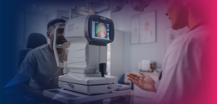
These imaging technologies can help with assessing unexpected structural and vascular retinal and choroidal changes in active and post-treatment toxoplasmic retinochoroiditis.

These imaging technologies can help with assessing unexpected structural and vascular retinal and choroidal changes in active and post-treatment toxoplasmic retinochoroiditis.

A study of adult patients with diabetes found that bowtie-shaped polarization artifacts do not serve as reliable clinical biomarkers for center-involving diabetic macular edema. What might this mean for clinicians?

Fundus autofluorescence is a helpful tool for diagnosing central serous choroidopathy and planning treatment.
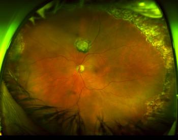
Physicians can discover, diagnose, document, treat diseases that might go undetected
Researchers seeking fluid features, retinal integrity measures to test next step in image analysis and image interrogation in clinical trials.
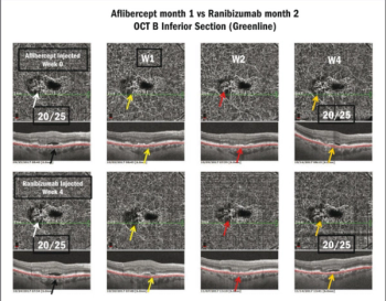
When used one week after injection, optical coherence tomography angiography can help clinicians determine how effective their treatment has been on neovascular membrane reperfusion.