













The investigators performed clinical examinations and obtained fundus photographs and inferior optical coherence tomography (OCT) images for both treatments at baseline and on days 1, 3, 8, 15, and 56 after the injections.

The investigators performed clinical examinations and obtained fundus photographs and inferior optical coherence tomography (OCT) images for both treatments at baseline and on days 1, 3, 8, 15, and 56 after the injections.

Researchers at the Warsaw, Poland-based International Center for Translational Eye Research decided to change that by introducing a new imaging method derived from optical coherence tomography. This led to the creation of even more advanced spatio-temporal optical coherence tomography.

The collaboration will drive an effort to develop predictive analytics to enable early detection of eye diseases and more personalized treatments to prevent vision loss for people with serious eye diseases.

Altris also has advanced AI models to enable vendor-neutral retina layers segmentation as well as detection and visualization of more than 70 retina conditions such as dry and wet age-related macular degeneration, geographic atrophy, diabetic retinopathy, diabetic macular edema and central retinal vein occlusion.
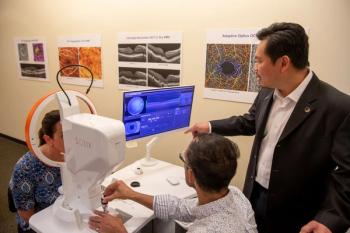
The Lasker-DeBakey Clinical Medical Research Award, dubbed "America’s Nobel," recognizes the wide use of optical coherence tomography to manage eye disease, prevent blindness.

Rather than speculating in a vacuum about the future of remote monitoring in ophthalmology, it is better to explore how other specialties in medicine adopted and integrated remote monitoring into their care plan frameworks.
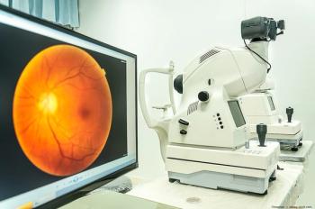
The company has released a video presentation that highlights combining genetic markers with high-resolution retinal imaging to assist in the detection and treatment of blindness and multiple systemic diseases.
The non-profit will partner with the imaging company to prevent blindness and fund new retinoblastoma research.

Optical coherence tomography analysis of measurements of cystoid macular lesions showed that patients with different IRD phenotypes can develop very large CMLs.
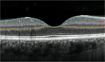
Researchers have identified markers that indicate the presence of Parkinson’s disease in patients an average of 7 years before the disease presents clinically.

According to Orbis International, the technology proves to be a practical solution in low-resource communities for tackling the leading cause of vision loss among working-age adults.

An experienced dermatologist blinded to clinical data performed the NVC using a videocapillaroscope VideoCap 3.0 (DSMedica) at 200× magnification in the study.

Shawn Kavoussi, MD, shared insights from his research on using infrared video for vitreous opacities and comparing postoperative reduction in vitreous opacity scores using infrared video with the Heidelberg spectralis unit.

The companies plant to create a comprehensive resource that provides valuable insights into health outcomes, medication adherence, and the effectiveness of various treatment protocols.

Research has found a new approach that identifies high-risk infants, so that not all preemies need to undergo an invasive eye exam.
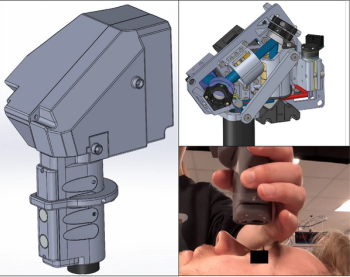
The investigators have developed a clinically robust HH-SECTR system, which has a modular aluminum probe design and micrometer-scale mechanical stability.
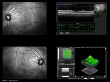
A single, consistent modality leads to better geographic atrophy tracking.

The retina is a peripheral part of the central nervous system and shares many similarities with cerebral brain matter.
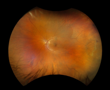
Optos announced the first ultra-widefield color rgb (red/green/blue) image. This new image modality is captured simultaneous to the optomap color rg image.

This medical device is the first retinal eye-movement monitor for non-invasive, objective clinical assessments.

Dierck Hillmann, PhD, was interviewed by our onsite team at the 2023 ARVO annual meeting where he shared research on holographic optical coherence tomography (OCT).

At the 2023 ARVO annual meeting in New Orleans, our on-site team caught up with T. Y. Alvin Liu, MD, to discuss predictive AI and its application in ophthalmology and screening of the eyes.

David Szanto, a medical student at Stony Brook, and working with the Department of Neurology at the Icahn School of Medicine at Mount Sinai, describes how an algorithm finds recurring patterns in the visual fields and the ganglion cell-inner plexiform layer thicknesses, and helps clinicians understand how prevalent those patterns are throughout the entire data.

The various aspects of home monitoring for patients with age-related macular degeneration are discussed in this point/counterpoint by Prof. Rufino Silva, University of Coimbra / Coimbra Hospital and University Centre, Portugal, and Univ.-Prof. Dr. med. Robert P. Finger, PhD, Department of Ophthalmology, University of Bonn, Germany, during the 14th annual Congress on Controversies in Ophthalmology in Lisbon, Portugal.

Imaging biomarkers are useful to determine the degree of EZ attenuation in the fovea.