





Imaging
Latest News
Advertisement
Video Series






CME Content











Advertisement
The biomarkers are especially crucial for patients with lupus nephritis.

The OcuMet Beacon is a proprietary, noninvasive tool for assessing retinal health. The company said it will be available on the US market for use in clinics and healthcare institutions.
The authors examined vascular and neural structures in the eye, assessing the foveal avascular zone and choroidal thickness

The approval makes ILM-Blue the first DORC posterior dye product available in China, according to a press release from Zeiss

One intravitreal injection of the dexamethasone implant in patients with refractory DME induced significant anatomic and functional improvements, but these improvements only lasted for short periods of up to 4 months.

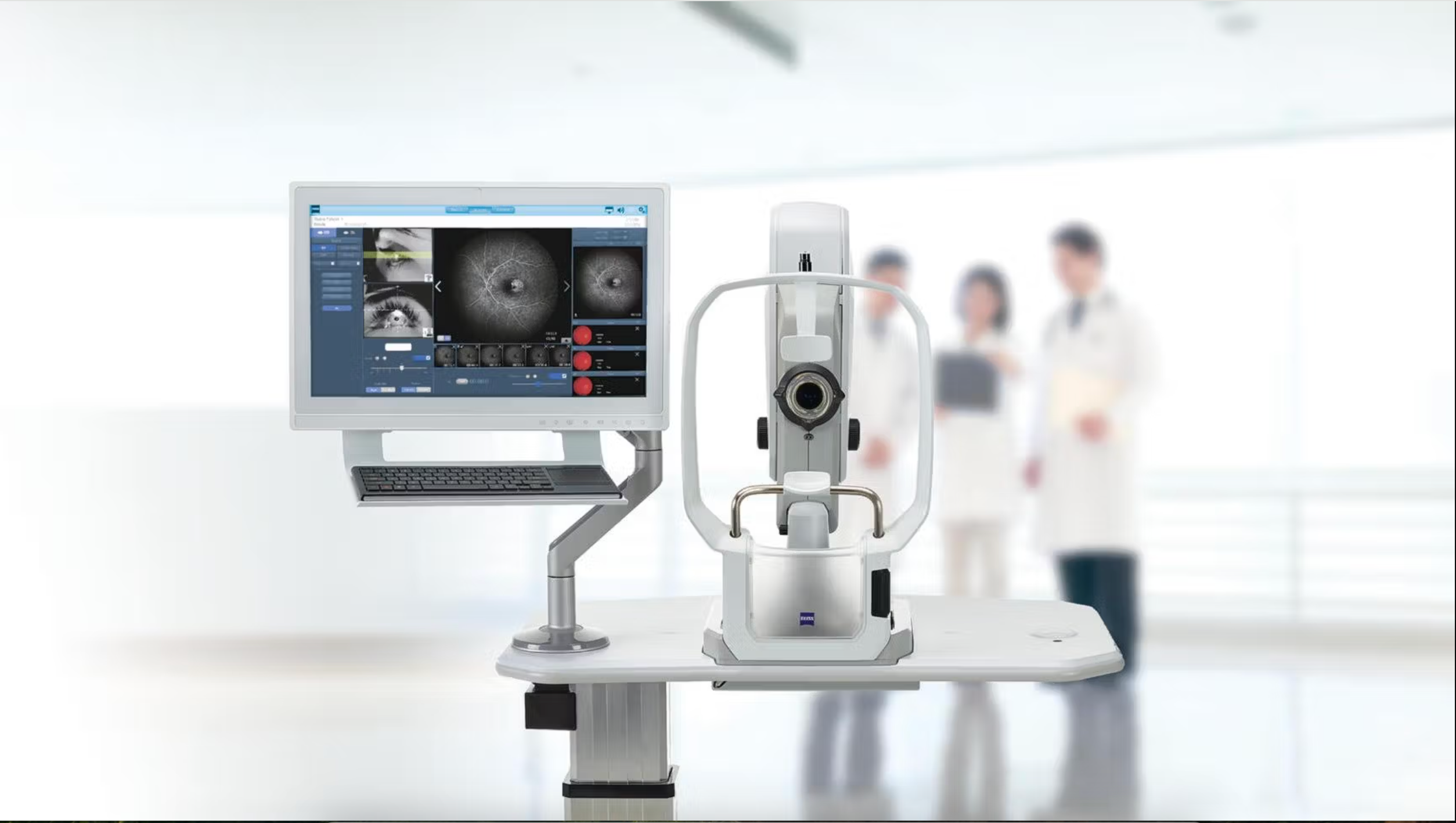
ZEISS VISULAS Combi is an advanced therapeutic laser workstation offering photodisruption, photocoagulation, and slit lamp technology in a single, comprehensive solution.
Systemic evaluation key to the cause of the detachments

Yu reviewed the benefits and limitations of various electrophysiologic technologies in a diverse group of inflammatory conditions affecting the retina and optic nerve.

The event will include presentations on artificial intelligence, ocular imaging and surgical topics.
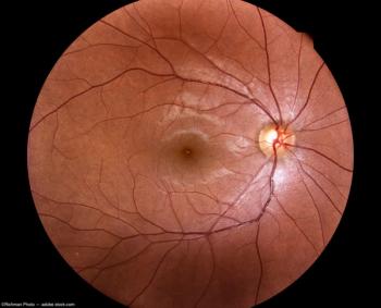
The imaging system is designed to enable secure, real-time remote monitoring around the clock, from virtually any location.
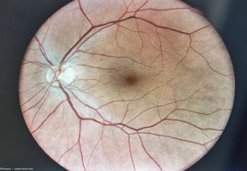
MonacoPro, the next evolution of Monaco from Optos, retains the powerful ultra-widefield SLO and spectral domain imaging while adding additional key product features.

Working definition forwarded by NIH CVI Working Group


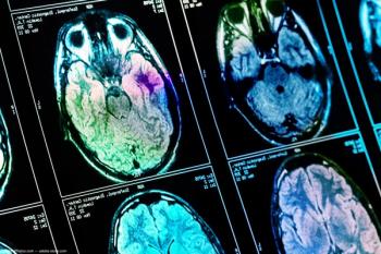
Researchers reported identifying changes in 4 areas of the brain

Optical coherence tomography angiography (OCTA) was performed in all participants; the choroidal and retinal changes were examined and recorded.
The “Triangle sign” seen on ultrasound is a “distinctive and reliable” ultrasound feature for differentiating total choroidal detachments and suprachoroidal hemorrhages.


The primary safety endpoint was carried out through the percentage of patients with shift from normal (at baseline) to abnormal in any electrocardiogram (ECG).
The findings suggest that retinal microvessel damage may occur due to renal hypertension (RH).
Clinical associations, imaging strategies and establishing a differential diagnosis from pseudopapilledema.
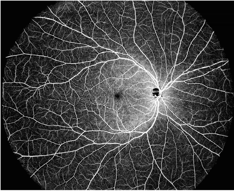
Peking University researchers have developed a deep learning-based, noninvasive choroidal angiography method that enables detailed 3D visualization of choroidal vessels from OCT scans. This technique could improve diagnostics for retinal diseases like macular degeneration, offering a safer alternative to traditional methods.
Advertisement
Advertisement
Trending on Eye Care Network - Ophthalmology Times
1
FDA approves phentolamine ophthalmic solution 0.75% (Ryzumvi) for presbyopia
2
Alvotech files first known aflibercept 8 mg biosimilar with EMA
3
Uveitis: a practical guide for ophthalmologists and optometrists
4
AAO 2026: How to become an ocular pain detective in dry eye
5