


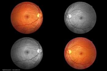
OCT
Latest News
Advertisement
Latest Videos

CME Content


Advertisement
More News
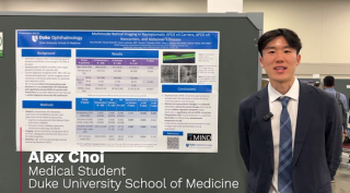
Celebrating a half-century of progress with Ophthalmology Times.
The investigators conducted a prospective interventional study to determine the therapeutic anatomic and functional effects of one intravitreal dexamethasone implant in eyes with refractory diabetic macular edema.

The Perimeter B-Series OCT system combines proprietary AI technology with optical coherence tomography, for use during breast-conserving surgeries.
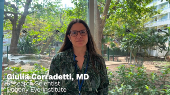
At the Envision Summit 2025 in San Juan, Puerto Rico, Giulia Corradetti, MD discussed AI applications in the identification and prediction of OCT structural biomarkers in intermediate AMD.
MonacoPro, the next evolution of Monaco from Optos, retains the powerful ultra-widefield SLO and spectral domain imaging while adding additional key product features.

Results from the study assisted Notal Vision in receiving De Novo authorization from the US FDA for the SCANLY Home OCT in 2024.


The primary safety endpoint was carried out through the percentage of patients with shift from normal (at baseline) to abnormal in any electrocardiogram (ECG).
Patients with fibromyalgia experienced changes in the macular ganglion cell layer and retinal nerve fiber layer.

With cases of syphilitic uveitis on the rise, recognizing its varied presentations—including optic disc edema, which may appear without other significant eye inflammation—is increasingly important for timely diagnosis and treatment.

Perimeter Medical Imaging AI has been leveraging the technology to improve breast cancer surgeries and reduce the number of patients requiring repeat surgery.
A study suggests that small hyperreflective retinal foci (HRF) on OCT images, linked to activated microglial cells, could serve as a valuable marker for monitoring neuroretinal inflammation and progression in age-related macular degeneration.

The NHS is expanding the use of optical coherence tomography to enhance diabetic eye care, aiming to improve early detection of diabetic retinopathy and reduce the backlog of hospital appointments.

News
A recent cohort study revealed low adoption of FDA-approved AI-based diabetic retinopathy detection, with less than 5% of diabetic patients receiving ophthalmic imaging. Researchers emphasize the need for improved awareness, cost-effectiveness, and integration to increase diabetic retinopathy screening rates.
News
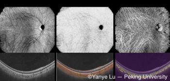
Peking University researchers have developed a deep learning-based, noninvasive choroidal angiography method that enables detailed 3D visualization of choroidal vessels from OCT scans. This technique could improve diagnostics for retinal diseases like macular degeneration, offering a safer alternative to traditional methods.

Researchers find a strong link between Alzheimer's disease and retinal thinning

News
In a study, a team of UCLA investigators detail a deep-learning model pre-trained on 2D scans that accurately predicts disease-risk factors from 3D medical-scan modalities.

News
The award from the National Institutes of Health will enable a team of researchers to investigate Alzheimer and Parkinson progression through the eye.
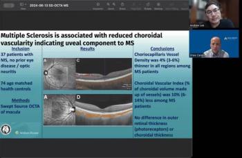
Andrew Lee, MD, and Andrew Carey, MD, sit down on another episode of the NeuroOp Guru to discuss Multiple Sclerosis and its association with reduced choroidal vascularity.

Ophthalmologists can use OCT to find the early signs of disease

The study helped identify predisposing factors for severe SANS in astronauts.

A team of researchers in China examined the difference and relationship between optical coherence tomography and optical quality analysis system parameters induced by compound electrolyte intraocular irrigating solution or Ringer lactate solution during uncomplicated cataract surgery.
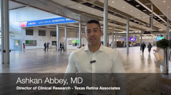
Ashkan Abbey, MD, spoke about his presentation on the CALM registry study, the 36-month outcomes of real-world patients receiving fluocinolone acetonide 0.18 mg at the annual ASRS meeting in Stockholm, Sweden.

The nOCT allows for detailed visualization of the brain's vascular anatomy.
Advertisement
Advertisement
Trending on Ophthalmology Times - Clinical Insights for Eye Specialists
1
Phase 3 NORTHSTAR trial launches for gildeuretinol in stargardt disease
2
Rising Star: Introducing Viha Vig, MBChB
3
Self-screening for ocular surface malignancies using a smartphone
4
Everest medicines acquires greater china rights to aceclidine presbyopia eye drop (VIZZ)
5