Publication
Article
Digital Edition
Are posterior corneal curvature changes culprit in hyperopic shifts?
Author(s):
Changes after DMEK, UT-DSAEK may be factors in reduced total corneal refractive power


Total corneal refractive power has always been a consideration, but not much has been known until now about why it changes in response to Descemet’s membrane endothelial keratoplasty (DMET) and ultrathin Descemet’s endothelial keratoplasty (UT-DSAEK).
The randomly selected, controlled Descemet Endothelial Thickness Comparison (DETECT) Trial identified decreases in the total corneal refractive power initially and then increases at 6 and 12 months postoperatively.
Related: Teaching AI algorithms to identify corneal pathology: The future is now
The changes seemed to reflect steepening in the posterior corneal curvature; despite that increase, the total corneal refractive power decreased from baseline to 12 months.
This may have implications for IOLs during cataract surgery, according to Elizabeth Shen, MD, of the Oregon Health and Science University in Portland.
To collect data on how the corneal refractive power responds to these corneal procedures, 50 eyes of 38 patients were randomly selected to undergo either DMEK or UT-DSAEK to address isolated endothelial dysfunction.
Tomography data were collected at baseline and postoperatively at 3, 6, 12, and 24 months.
The primary outcomes of the DETECT Trial were the total corneal refractive power at the 3-mm zone, anterior and posterior simulated keratometry, and the predicted refractive outcome using the Holladay 1 formula compared with the postoperative manifest refraction.
Investigators also evaluated patients who underwent endothelial keratoplasty and cataract surgery.
Related: Perioperative medications can eliminate postoperative drops after cataract surgery
Findings
The total corneal refractive power decreased in response to both DMEK and UT-DSAEK at 3 months postoperatively and then increased at 6 and 12 months.
Despite the tendency to increase, the parameter overall decreased significantly (P < .001 for all comparisons at each time point) with both procedures from baseline to 24 months, Shen reported.
The mean change in the total cornea refractive power after DMEK from baseline to 12 months was a reduction of + 0.8 dD and for UT-DSAEK a reduction of + 0.69 D. The difference did not reach significance.
This raised the question of why a decrease in the total corneal refractive power occurs. When investigators evaluated the anterior corneal curvature in relation to the changes from baseline out to 12 months, they found that the change with DMEK was + 0.23 D and with UT-DSAEK + 0.11, not significant from baseline.
Related: Hardware, software offer surgeons a window to cornea diagnosis
The mean change in the posterior corneal curvature at 12 months with DMEK was – 0.42 D and with UT-DSAEK – 0.54 D, which also was not significant.
Shen noted that the decrease represents a steepening of the posterior corneal curvature. At each time point evaluated, there was a significant change compared with baseline.
The investigators also looked at the refractive outcomes after triple procedures. The analysis showed a mean refractive shift toward hyperopia of + 0.6 D with DMEK and with UT-DSAEK + 0.4 D.
Most of the outcomes had a hyperopic shift from the so-called triple procedures, but a myopic shift also occurred. No difference was seen in the refractive outcomes between DMEK and UT-DSAEK.
Related: ASCRS 2020: Returning to baseline post-corneal collagen crosslinking for keratoconus
A key takeaway point is that the total corneal refractive power in DMEK and UT-DSAEK decreased at first and then increased at 6 and 12 months.
Despite the increase, the total corneal refractive power decreased from baseline to 12 months.
This loss of corneal power likely results from changes in the posterior curvature, which steepened over time and contributed to hyperopic shifts after endothelial keratoplasty.
The results did not differ between DMEK and UT-DSAEK.
--
Elizabeth Shen, MD
e: shene@ohsu.edu
Shen has no financial interest in this subject matter.


Newsletter
Don’t miss out—get Ophthalmology Times updates on the latest clinical advancements and expert interviews, straight to your inbox.
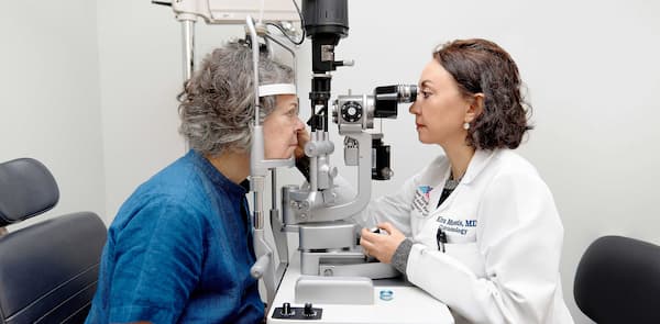
 Inside NYEE’s new refractive solutions center with Kira Manusis, MD")

 ASCRS 2025: Eric D. Donnenfeld, MD, on the effect of lifitegrast clinical signs and biomarkers in dry eye disease")
 ASCRS 2025: Amar Shah, MD, on why hyperosmolar tear film before and after cataract surgery matters")


 ASCRS 2025: Jason Bacharach, MD, on early-onset efficacy with perfluorohexyloctane in dry eye")
 ASCRS 2025: Karl Stonecipher, MD, on LASIK outcomes using an aspheric excimer laser for high myopia")
 ASCRS 2025: Joaquin De Rojas, MD, leverages machine learning model to predict arcuate outcomes")