

|Articles|August 24, 2021
Corneal keratometry and corneal astigmatism changes after surgery for acquired ptosis
Author(s)Lynda Charters
Researchers conducted a retrospective observational study to assess the corneal refractive changes induced by surgery to correct ptosis using Fourier harmonic analysis.
Advertisement
Significant changes were seen in the anterior and posterior corneal keratometry and posterior corneal astigmatism 6 months after ptosis surgery performed to correct acquired ptosis, according to first author Risako Yamamoto, MD, from the Department of Ophthalmology, Graduate School of Medicine, the University of Tokyo, Tokyo, Japan.
She and colleagues from the Department of Ophthalmology, Keio University Hospital, Tokyo, conducted a retrospective observational study1 to assess the corneal refractive changes induced by surgery to correct ptosis using Fourier harmonic analysis.
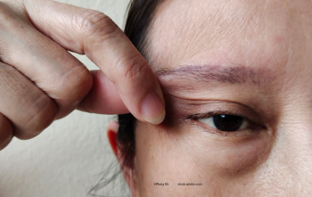
Consecutive patients were enrolled who had undergone levator aponeurotic surgery for acquired ptosis from May 2016 to January 2018. The surgeries were performed at the University of Tokyo Hospital.
The investigators analyzed the best-corrected visual acuity (BCVA), central corneal thickness (CCT), average, corneal astigmatism, and topographic data using Fourier analysis preoperatively and 6 months postoperatively.
A total of 32 eyes of 32 patients (mean age, 72.6 ± 8.5 years) were included. The investigators reported that no significant differences were found in the BCVA and the CCT.
They did, however, find significant decreases in the anterior average keratometric corneal power, anterior corneal astigmatism, and posterior corneal astigmatism 6 months postoperatively (p < 0.001 for all comparisons).
Fourier harmonic analysis showed that the anterior spherical component decreased significantly (p < 0.001) at the 6-month time point.
The other parameters of the anterior and posterior cornea did not differ significantly.
Significant negative correlations were seen between the preoperative posterior average keratometric corneal power and the changes in the posterior average keratometric corneal power (r = −0.891, p < 0.001) and between the preoperative posterior corneal astigmatism and the changes in the posterior corneal astigmatism 6 months postoperatively (r = −0.858, p < 0.001), the investigators reported.
They concluded that the anterior and posterior keratometry and posterior corneal astigmatism changed significantly as a result of the surgery to correct acquired ptosis.
Reference
- Yamamoto R, Ono T, Toyono T, et al. Assessment of long-term anterior and posterior topographic changes in the cornea after ptosis surgery using Fourier harmonic analysis. Cornea 2021;40:440-444; doi: 10.1097/ICO.0000000000002429
Advertisement
Related Content
Advertisement


Latest CME


Advertisement
Advertisement