


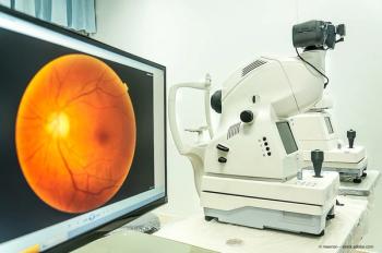
OCT
Latest News
Advertisement
Latest Videos
Shorts
1:36
Ophthalmology without OCT - Part 5
3 months ago
by
David A. Eichenbaum, MD, FASRS(+2 more)

1:20
Ophthalmology without OCT - Part 4
3 months ago
by
Nimesh Patel, MD(+3 more)
1:12
Ophthalmology without OCT - Part 3
3 months ago
by
Michael Lai, MD, PhD(+2 more)
1:24
Ophthalmology without OCT - Part 2
3 months ago
by
Justis P. Ehlers, MD(+2 more)
1:17
Ophthalmology without OCT
3 months ago
by
Andrew J. Barkmeier, MD(+3 more)
CME Content


Advertisement
More News
As ophthalmic technologies move at supersonic speed, AI and gene therapy take center stage.
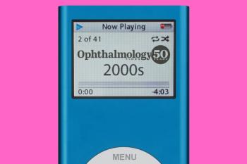
Explore groundbreaking advancements in eye care from the 2000s, including LASIK innovations, anti-VEGF treatments, and enhanced imaging technologies.
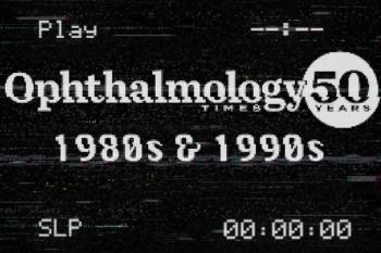
Explore the groundbreaking advancements in ophthalmic technology from the 1980s and 1990s, including excimer lasers, LASIK, and innovative imaging techniques.

Conavi leverages ophthalmic imaging technology to enhance intravascular diagnostics in cardiology.

Recapping the Heidelberg Engineering International SPECTRALIS Symposium—and Beyond.

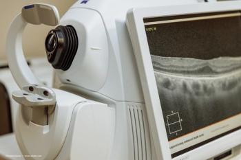
Advanced imaging and awareness of systemic risk factors are essential.

Ophthalmology celebrates 50 years of groundbreaking innovations, highlighting anti-VEGF therapies, OCT imaging, and future advancements in eye care.

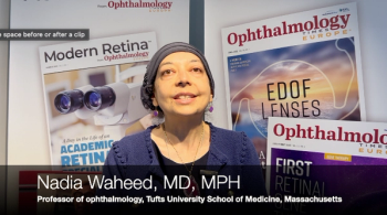
To mark Ophthalmology Times' 50th anniversary, we invited top experts to reflect on the most significant innovations in ophthalmology over the past five decades.
To mark Ophthalmology Times' 50th anniversary, we invited top experts to reflect on the most significant innovations in ophthalmology over the past five decades.

ZinserLab will focus on accelerating the development of imaging technologies in eye care.
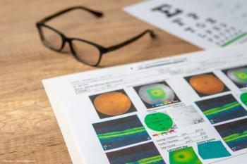
The investigators described the CASIA2 (Tomey Corporation) instrument, which is a new AS-OCT device with a swept-source laser wavelength of 1,310 nm that can scan at a speed of 50,000 A-scan/second.
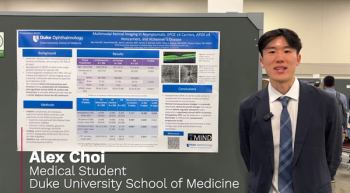
At ARVO 2025, in Salt Lake City, Utah, Alex Choi discussed his poster on APOE carrier genetic status and its relation to Alzheimer disease as well as changes of the retina that might reflect early signs leading to an earlier diagnosis of the disease

Join global experts at the 2025 International SPECTRALIS Symposium in Heidelberg, exploring innovations in ophthalmology and space-related ocular health.
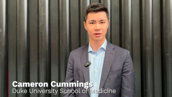
At the ARVO 2025 meeting in Salt Lake City, Utah, Cameron Cummings presented a poster on how retinal microvascular imaging metrics may show sex-specific biomarkers for Parkinson's disease.

Brazilian researchers reveal how choroidal thickness influences treatment response in diabetic macular edema, enhancing personalized therapy strategies.
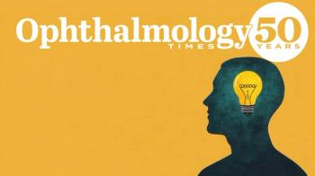
Celebrating a half-century of progress with Ophthalmology Times.
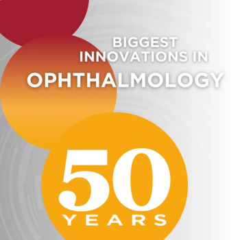
In celebration of Ophthalmology Times' 50th anniversary, we asked leading experts in the field what they see as the biggest innovation in ophthalmology in the past 5 decades.
In celebration of Ophthalmology Times 50th anniversary, we asked leading experts in the field what they see as the biggest innovation in ophthalmology in the last 5 decades.
In celebration of Ophthalmology Times 50th anniversary, we asked leading experts in the field what they see as the biggest innovation in ophthalmology in the last 5 decades.
In celebration of Ophthalmology Times 50th anniversary, we asked leading experts in the field what they see as the biggest innovation in ophthalmology in the last 5 decades.
The investigators conducted a prospective interventional study to determine the therapeutic anatomic and functional effects of one intravitreal dexamethasone implant in eyes with refractory diabetic macular edema.
Advertisement
Advertisement