

|Articles|July 26, 2019
Photoreceptor replacement cells enter therapeutic space
Author(s)Lynda Charters
Ganglion, bipolar cells increase options for patients with late-stage retinal disease
Advertisement
Treating late-stage retinal disease with induced pluripotent stem cells is on the clinical horizon.
Reviewed by Edwin M. Stone, MD, PhD
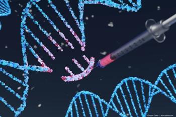
Human retinal engineering is a rapidly growing field, albeit an expensive one.
“Cost containment is becoming increasingly important in medicine because without some type of substantial governmental or actuarial wealth redistribution, the vast majority of people in the United States will not be able to afford the life-changing treatments that science is developing,” said
Goals of stem cell transplantation
The goal of this technology is durable restoration of useful vision in patients with advanced stages of retinal degeneration. These diseases range from very common
“What unifies these conditions is the selective loss of the outer retina,” said Dr. Stone, professor of ophthalmology, and the director of the
The key players involved with vision in the retina are the photoreceptors, bipolar cells, and ganglion cells. In AMD and RP, the photoreceptors are lost, but the ganglion and bipolar cells remain over decades even in the presence of no light perception vision, he explained. Recognition of this has set the stage for transplantation of a graft into the subretinal space to replace the photoreceptors.
Dr. Stone noted that this has already been achieved in a few different animal models. In a blind mouse model, increased electroretinographic activity was observed with increasing doses of injected cells.
How it works
When considering this type of intervention in humans, Dr. Stone pointed out the need for two strategic decisions: first, whether to use autologous cells or cells from another individual and, second, the type of injection, i.e., a bolus injection or a polymer supported sheet. In Dr. Stone’s group, the investigators have opted to use autologous cells that are supported by a polymer.
They reached this decision, he explained, because of the increasing evidence that retinal allografts are associated with a robust immune response even when the recipient has a normal retina before transplantation.
Autologous grafts have the added advantage that they will minimize the need for expensive, somewhat toxic, life-long immunosuppression.
RELATED:
“If new retinal cells can be derived from the patient for whom they are intended, those cells would be the best possible immunologic match and require the least immunomodulation,” he explained.
Induced pluripotent stem cells (iPSCs) can be differentiated into photoreceptor precursor cells for autologous transplantation. However, the downside to this strategy is that in a patient with RP they still contain the mutations that caused the disease.
To counteract this, the
Photoreceptor mechanisms
Dr. Stone explained the procedure to create patient-derived photoreceptor precursor cells. The first step is obtaining a skin biopsy in order to establish a fibroblast culture.
These fibroblasts are then treated with four pluripotency factors that erase the cells differentiated state and then turn them into iPSCs. When cultured with additional growth factors that drive core brain development, the cells begin developing structures that resemble eye cups after about 30 days.
The eye cups then are removed and cultured with growth factors that induce further retinal development. “In a matter of weeks, transplantable photoreceptor precursor cells are available,” Dr. Stone stated.
However, the vast majority of the cells, i.e., 90%, will die if they are just injected as an unsupported bolus under the retina. This is the stage at which the polymer support, which can be created using a three-dimensional printer, comes into play. The polymer support increases the cellular survival rate.
The printer can print dissolvable biopolymer scaffolds at a subcellular scale, he explained. The scaffold has three-dimensional channels that are about 10 microns in diameter. In the rat, optical coherence tomography showed an intact retina draped over a graft that was slowly dissolving 6 months after implantation.
The first trial performed in completely blind patients will involve implantation of a 5-mm circular graft containing about 500,000
To make this technology available throughout the country, multiple regional stem cell facilities connected to surgical suites will need to be established because the grafts are living tissue that cannot be transported long distances.
“Photoreceptor replacement therapy is steadily moving closer to the clinic,” Dr. Stone concluded. “With the use of patient-derived cells, we can reduce the need for expensive long-term immunosuppression. Robots will help reduce manufacturing costs, which will increase access to these treatments for our patients.”
RELATED:
Disclosures:
Edwin M. Stone, MD, PhD
E: [email protected]
Dr. Stone has no financial interest in any aspect of this report.
Newsletter
Don’t miss out—get Ophthalmology Times updates on the latest clinical advancements and expert interviews, straight to your inbox.
Advertisement
Related Content
Advertisement


Latest CME


Advertisement
Advertisement
Trending on Ophthalmology Times - Clinical Insights for Eye Specialists
1
MeiraGTx Licenses complement-targeted geographic atrophy program from ZipBio
2
Metformin use associated with reduced incidence of intermediate AMD
3
Last year in glaucoma at EnVision Summit 2025
4
Looking back at the 2025 EnVision Summit
5