Thermography useful in evaluating filtering bleb function
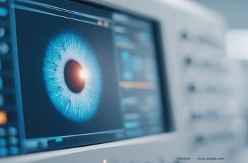
Because the temperature decrease in the filtering bleb after trabeculectomy provides information about its function, a new ocular surface-oriented, infrared radiation thermographic device (TG 1000, Tomey) may help evaluate filtering bleb function. The device is easy to handle and creates reproducible measurements, according to new research published in the Journal of Glaucoma.
Because the temperature decrease in the filtering bleb after trabeculectomy provides information about its function, a new ocular surface-oriented, infrared radiation thermographic device (TG 1000, Tomey) may help evaluate filtering bleb function. The device is easy to handle and creates reproducible measurements, according to new research published in the
Researchers prospectively studied one eye in each of 35 patients after the eyes had undergone trabeculectomy. They tested the functioning of the filtering bleb using the new device in a noncontact manner. The investigators used patients’ postoperative IOP to classify the eyes as having IOP that was either poorly controlled or well-controlled.
To evaluate bleb function, the researchers used the mean temperature decrease in the filtering bleb (TDB); TDB=(mean temperature of the temporal and nasal bulbar conjunctiva)−(mean temperature of the filtering bleb). They evaluated the filtering bleb during 10 seconds of eye opening and also introduced a new parameter: the TB10sec.
In the well-controlled IOP group, the authors found that the TDB was 0.911° C and the TB10sec was −1.027° C. In the poorly controlled IOP group, the TDB was 0.599° C and the TB10sec was −0.623° C. These differences were significant.
Newsletter
Don’t miss out—get Ophthalmology Times updates on the latest clinical advancements and expert interviews, straight to your inbox.


