News
Article
CU Ophthalmology residents employ portable fundus photography cameras to enhance on-call imaging
Author(s):
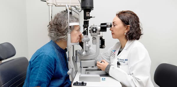
Mobile cameras allow University of Colorado ophthalmology residents working in hospital settings to conduct more comprehensive and efficient patient evaluations.

")
(Image Credit: AdobeStock/heitipaves)
For ophthalmology residents at the University of Colorado School of Medicine, working on-call in a hospital can lead to a mixed bag of eye or vision complaints and they have to be ready for anything.
Portable fundus photography cameras are giving them cutting-edge tools at their fingertips.
“Ophthalmology is unique because it’s a clinic-based specialty, and we rely on specialized equipment to get the most accurate diagnosis and treatment plan,” third-year resident Nihaal Mehta, MD, said in a news release.1 “We have a backpack stocked with dozens of eyedrops and tools that help us perform even seemingly simple tasks, like checking eye pressure, for example.”
The Colorado University Department of Ophthalmology added the portable cameras more than a year ago to the backpacks of residents working on-call, kicking off a new era for both resident training and patient care.
According to the news release, the portable cameras are about the size of a smartphone and are equipped with a long lens that brings valuable imaging to wherever a resident is needed. The addition to these cameras, the university noted, has boosted the first evaluation of patients and facilitated follow-up care. Supervising physicians can now easily see what a resident is seeing, confirming a treatment plan or being able to access more information when a question or concern arises.1
UC professor and department chair Naresh Mandava, MD, pointed out that the technology offers another option during diagnosis and treatment.
“As a retina specialist, I have always been fascinated by the value of an ophthalmic image, not only in making a diagnosis but also in sharing cases with colleagues,” Mandava said in the news release. “The opportunity to deploy a simple imaging technology into a dynamic tertiary care center environment has the potential to improve outcomes for patients as well as decrease the time needed for consultation with colleagues and other experts.”
A new use for a trusted tool
According to the university, Phelcom gifted its department 3 Eyer portable cameras which enable residents to have them on hand where they may be scheduled to work. This can save them time, particularly in fast-paced situations.
At UCHealth University of Colorado Hospital, on-call residents completed 1200 emergency department consults and 800 inpatient consults in 2023. They also cross the UC Anschutz campus to see patients at the Rocky Mountain Regional VA Medical Center and Children’s Hospital Colorado.1
“Residents might see 10 to 15 patients in 24 hours, so it can be really hectic,” Mehta said in the news release. “Any tool that can help you while you’re on-call is a huge godsend.”
Moreover, the university noted that eye pathology can be especially difficult to put into words, making these new cameras all the more helpful. The images have increased communication between residents, fellows, and supporting doctors, who might’ve previously needed to re-evaluate a patient for a second time because they only had a written description of the condition.1

")
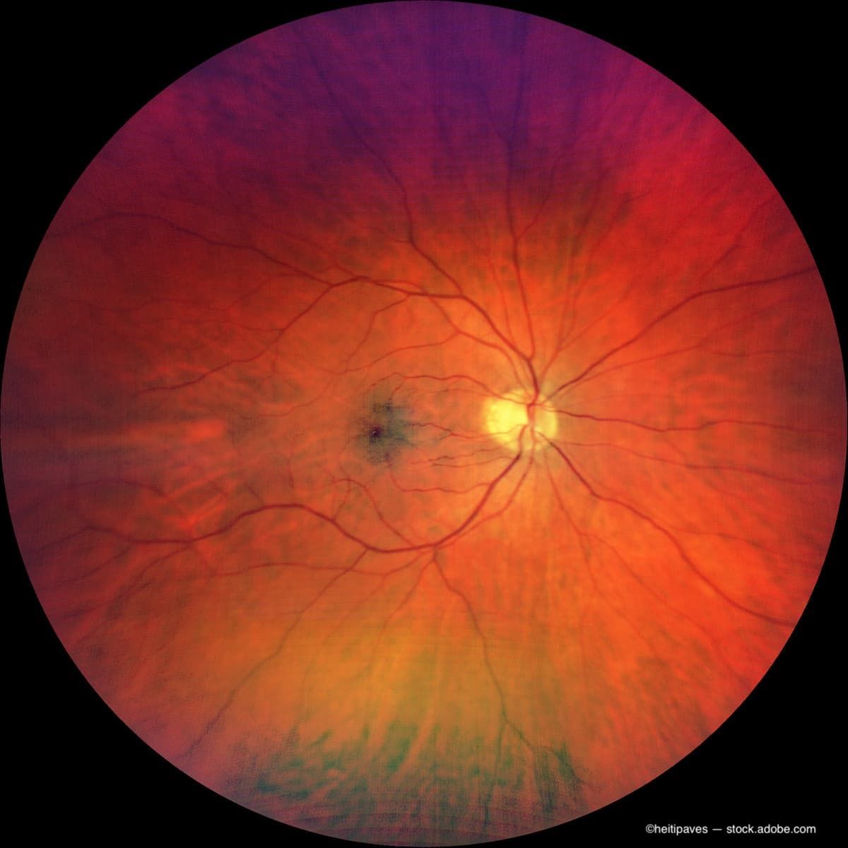
An image from a portable fundus photography camera used by CU ophthalmology residents shows severe papilledema from malignant idiopathic intracranial hypertension. The images became the basis for a paper resident Nihaal Mehta, MD, and faculty published in October 2022. (Image credit: Colorado University Department of Ophthalmology)
“These cameras expedite the process and make it more streamlined when verifying exam findings,” Mehta says. “It helps us to make a clinical plan, which is also really helpful for the patient."
The cameras are proving to be a handy tool for resdients in the long term, too.
“These cameras have been helpful in monitoring patients over time,” Mehta pointed out in the news release “If a patient has bleeding in the back of their eye, you can describe what you see, but a photo a week later can tell you more accurately how the condition is changing.”
In the classroom
Residents also are finding the tools useful outside of the hospital setting, including in classroom and academic situations.
“This helps us to present at a conference or publish a paper,” Mehta explained. He was able to use an image taken with a portable fundus camera to help finish a paper on a rare exam finding.2
“Because we had those photos, we were able to submit it as a picture case,” he pointed out.
Mandava explained that the residents also bring photos from the camera to weekly conferences where they discuss cases.
“Every Wednesday morning, we have a debrief with all the residents on interesting cases from the past week. The addition of imaging to the consult workflows has improved the educational environment for our residents, fellows, and faculty alike,” Mandava concluded in the news release. “In addition, the time needed to gain multiple opinions has been reduced drastically, which is ultimately a benefit to the patients.”
Reference:
CU Ophthalmology residents employ portable fundus photography cameras to enhance on-call imaging. EurekAlert! Accessed February 29, 2024. https://www.eurekalert.org/news-releases/1035799
Anna Strong Caldwell, MD, Nihaal Mehta, MD, Prem S. Subramanian, MD, PhD. Malignant Idiopathic Intracranial Hypertension. Ophthalmology. Published October 10, 2022. Accessed February 28, 2024. DOI: https://doi.org/10.1016/j.ophtha.2022.09.006.
Newsletter
Don’t miss out—get Ophthalmology Times updates on the latest clinical advancements and expert interviews, straight to your inbox.
 NeuroOp Guru: Using OCT to forecast outcomes in ethambutol optic neuropathy")
 Anat Loewenstein, MD, shares insights on the real-world results of remote retinal imaging")
 FLIO and the brain: Making the invisible visible with Robert Sergott, MD")
 Structure-function correlates using high-res OCT images with Karl Csaky, MD, PhD")
 SriniVas Sadda, MD, on high-res OCT of atrophic and precursor lesions in AMD")
 Christine Curcio, PhD, shares histology update supporting review software and revised nomenclature for <3 μm OCT")


 NeuroOp Guru: When eye findings should prompt neuroimaging in suspected neuro-Behcet disease")