The gene therapy technology already exists to treat most inherited retinal disease. The current challenge is to drive down the costs of implementing the technology.

The gene therapy technology already exists to treat most inherited retinal disease. The current challenge is to drive down the costs of implementing the technology.

The FDA recently granted regulatory approval for tocilizumab (Actemra, Genentech), the first approved therapy for the treatment of adults with giant cell arteritis.

Endoillumination for vitreoretinal surgery continues to improve with the introduction of brighter, safer light sources with color filters that may allow opportunities for better tissue visualization.

Compounded pharmaceuticals have been used for decades, but does that mean they are reliable and safe? “Absolutely,” asserted Dante Pieramici, MD. “The literature disagrees,” said Julia Haller, MD.

Local therapy plays a significant role in the treatment of uveitis, either as monotherapy or in combination therapy. Alongside a number of pharmacotherapeutic agents, new products and technologies are in development that could expand clinicians’ options and improve outcomes.

The UK’s National Institute for Health and Care Excellence (NICE) has given preliminary approval to dexamethasone 0.7 mg intravitreal implant (Ozurdex, Allergan) and adalimumab (Humira, AbbVie) for sight-threatening non-infectious posterior uveitis.

The efficacy of intravitreal bevacizumab (Avastin, Genentech) for improving vision in patients with center-involving diabetic macular edema (DME) has been demonstrated in several clinical trials.

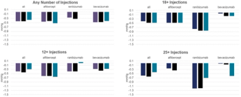
All three anti-vascular endothelial growth factor (anti-VEGF) agents that are commercially available have demonstrated efficacy for the treatment of diabetic macular edema (DME). However, it is clear from the results of clinical trials that outcomes with bevacizumab are not as good as those obtained using ranibizumab or aflibercept, according to Jean-François Korobelnik, MD.

Eyes operated on with pars plana vitrectomy needed fewer reoperations over 180 days than eyes subjected to scleral buckling (SB) in a retrospective comparison of patients with rhegmatogenous retinal detachment (RRD).

Evidence supports the involvement of an important pathway in retinal ganglion cell dysfunction and death in traumatic optic neuropathy.

Key components in retinopathy of prematurity trials include risk factors, predictive models, telemedicine and tele-education trends, and treatment.

A phase III study program investigating abicipar pegol (Allergan) for the treatment of neovascular age-related macular degeneration (nAMD) is underway based on promising efficacy and safety results in phase II studies.

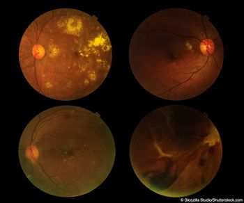
Multimodal imaging, including traditional and newer techniques, is necessary for identifying the spectrum of retinal lesions associated with age-related macular degeneration (AMD), said David Sarraf, MD.

Earlier this month, a jury convicted West Palm Beach retina surgeon Solomon Melgen, MD, on 67 counts of Medicare fraud. This high-profile case hit very close to home, as myself and another retina specialist from my own community were on opposing sides of the case.

Laser photocoagulation is a time-tested, well-defined, and effective treatment for retinopathy of prematurity (ROP) with requirements for follow-up that are much more manageable compared with anti-vascular endothelial growth factor (anti-VEGF) therapy, argued David K. Wallace, MD, MPH.

Anti-VEGF therapy for Zone I retinopathy of prematurity (ROP) is associated with better structural and functional outcomes than laser therapy, but also a longer recurrence risk period, said Helen A. Mintz-Hittner, MD.

I have been encouraged as a retinal specialist with all the excitement and promising new treatments in gene therapy. Given that there are several retinal diseases with known inherited patterns and confirmed genetic loci responsible for the clinical manifestations, our field is primed to benefit from the expansion of knowledge in this arena.

Intravitreal aflibercept worked better than panretinal photocoagulation (PRP) for proliferative diabetic retinopathy (PDR) in a new head-to-head trial, according to researchers.

Activation of Tie2–as a result of subcutaneous administration of AKB-9778 (Aerpio Therapeutics) in combination with an anti-vascular endothelial growth factor (anti-VEGF) therapy–enhances the effect of an anti-VEGF drug on diabetic macular edema (DME).

Analyses of data collected in the RISE/RIDE and VIVID/VISTA clinical trials provide important messages about the efficacy and safety of ranibizumab (Lucentis, Genentech) and aflibercept (Eylea, Regeneron) for treatment of diabetic macular edema (DME).

Endocryotherapy is a good treatment option for patients with larger retinal angiomatous tumors.

Nucleoside-reverse, transcriptase inhibitors (NRTIs) or their derivatives could one day be used to prevent or treat age-related macular degeneration (AMD), according to Jayakrishna Ambati, MD.

Results from the Study Assessing double-masKed Uveitis tREAtrement (SAKURA) program support the efficacy and safety of intravitreal sirolimus 440 mcg (Opsiria, Santen) for treating noninfectious uveitis of the posterior segment, said Pauline T. Merrill, MD.

Results from the Study Assessing double-masKed Uveitis tREAtrement (SAKURA) program support the efficacy and safety of intravitreal sirolimus 440 mcg (Opsiria, Santen) for treating noninfectious uveitis of the posterior segment, said Pauline T. Merrill, MD.

The Diabetic Retinopathy Clinical Research Network’s (DRCR.net) Protocol T-the first head-to-head-to-head comparison among aflibercept (Eylea, Renegeron Pharmaceuticals), ranibizumab (Lucentis, Genentech), and bevacizumab (Avastin, Genentech)-found in its first-year results that all three agents improved vision and reduced edema effectively.

The scientific community knows that vascular endothelial growth factor (VEGF) causes increased, vascular permeability, resulting in diabetic macular edema (DME) in the ischemic retina, but how to stop the VEGF drive remains the challenge.

Removing a small area of internal limiting membrane at the intended site of a subretinal injection allows the injection to be delivered without penetrating the retina.

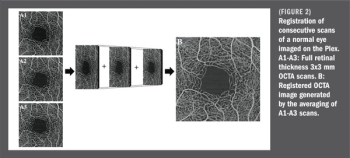
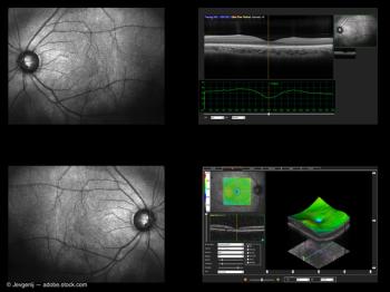
New imaging technologies, such as ultra-wide-field fluorescein angiography and optical coherence tomography angiography, are providing more details about retinal diseases than previous imaging technologies, leading to better understanding of disease processes.

Peeling of the internal limiting membrane with staining of retinal tissues might help macular hole closure and resolve retinal schisis.

Knowledge of pathologic patterns is necessary in medical retina, especially patterns that are unusual or rare. Pattern recognition skills are essential in medical retina. Lawrence Yannuzzi, MD, highlights four patterns that he believes are relevant in clinical practice and should be recognized–and not missed–by retinal specialists.