
News|Articles|March 18, 2019
- Ophthalmology Times, March 15 2019
- Volume 44
- Issue 5
Long-term data establish SMILE role in myopic treatment armamentarium
Author(s)Cheryl Guttman Krader, BS, Pharm
Follow-up at 7 years shows procedure safe, effective, provides stable correction
Advertisement
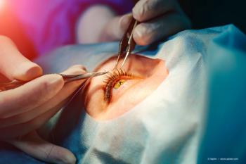
Follow-up that includes data from 264 eyes seen at 7 years after surgery show that small-incision lenticule extraction (SMILE, Carl Zeiss Meditec) is safe, effective, and provides stable correction for eyes with up to –14.0 D of myopia and up to –6.0 D of astigmatism.
Reviewed by Osama Ibrahim, MD, PhD
Results from long-term follow-up show that small-incision lenticule extraction (SMILE, Carl Zeiss Meditec) provides safe, effective, and stable correction of low to high myopia with astigmatism, said Osama Ibrahim, MD, PhD.
“We have been doing SMILE for more than 10 years, and there are still some challenges that need to be addressed, but SMILE is here to stay,” said Dr. Ibrahim, professor of ophthalmology, Alexandria University, Alexandria, Egypt. His comments were based on data from 5,263 eyes, of which 264 were seen after 7 years.
Because of access to research software, the amounts of sphere and cylinder corrections exceeded those available commercially. Preoperatively, manifest refraction spherical equivalent (MRSE) for the entire cohort averaged –5.92 D ± 2.13 D and ranged from –1.25 to –14.00 D; MR cylinder averaged –1.26 D ± 1.04 D and ranged up to –6 D. and we get very good results.
At 1 week after surgery, mean MRSE was –0.15 D ±0.51 D (range, +2.00 to –3.50 D) and mean MR cylinder was –0.12 ±0.38 D (range, 0.00 to –2.75 D).
“Some cases were initially undercorrected intentionally, and we had some overcorrections that occurred because there was no software adjustment at the time of the procedure,” Dr. Ibrahim said.
Data from follow-up at 1 year was available for 3,876 eyes, 587 eyes were seen at 2 years, and 264 eyes were seen at 7 years. Results from manifest refraction were generally stable over time except for a slight increase MRSE in later years.
“Many of the patients were treated when they were just 19 or 20 years old, and the late change in MRSE is explained by an increase in axial length over time,” Dr. Ibrahim said. “The MR cylinder, however, was unchanged.”
Refractive predictability was excellent for the overall cohort and across all subgroups when eyes were divided by degree of myopia into mild-moderate (MRSE <–6.0 D), high (MRSE ≥–6.0 to <–10.0 D) and very high (MRSE ≥–10.0 D). At different follow-up intervals, achieved MRSE was ±0.25 D of target in about 80% of eyes.
“SMILE is not a procedure for treating a specific level of myopia,” Dr. Ibrahim said. “Rather, it can treat a broad range.”
Analyses of changes from baseline BSCVA provided assurance about the safety of the procedure. The percentage of eyes losing 2 or more lines from preoperative BSCVA was ≤1% at any follow-up; BSCVA was unchanged or improved in 89% to 95% of eyes across the different follow-up intervals, and the percentage of eyes that gained 1 or more lines of BSCVA increased over time.
“Safety was our main concern when we started with SMILE because we were still refining the techniques and technology,” Dr. Ibrahim said. “Luckily, we found in our follow-up that some early losses of BSCVA were transient.”
He pointed out that gains in BSCVA after SMILE may be explained not just by loss of minification but by the flattening response that occurs after SMILE. Topographic maps show a 6.0- or 6.5-mm diameter area of flattening following removal of a 6.0-mm lenticule whereas after LASIK, the area of flattening is smaller in diameter than the ablation zone.
“The cornea responds differently to tissue removal than it does to ablation,” Dr. Ibrahim said. “The peripheral cornea relaxes after SMILE, and we believe that is why we can use SMILE to correct higher amounts of myopia and obtain better quality of vision compared with LASIK.”
Remaining issues
Correction of residual refractive error after SMILE has been done with LASIK and PRK, but ongoing research is investigating enhancement by performing SMILE in the cap or in the residual bed. So far, the latter techniques have been associated with encouraging results.
Other techniques under development include customization (wavefront guidance), cyclotorsion adjustment, and treatment of hyperopia. In addition, more study is needed to prove that SMILE has an advantage over LASIK when it comes to preserving corneal biomechanical integrity and to corroborate its long-term stability.
“Some people still need to be convinced that we are not getting regression after SMILE,” Dr. Ibrahim said. “Looking at eyes in our series that had residual error later during the follow-up we found that they had the same error from the beginning.”
Disclosures:
Osama Ibrahim, MD, PhD
E: [email protected]
This article was adapted from Dr. Ibrahim’s presentation during Refractive Surgery Subspecialty Day at the 2018 meeting of the American Academy of Ophthalmology. He is a consultant to Carl Zeiss Meditec.
Articles in this issue
over 7 years ago
Valuing power of the lenticleover 7 years ago
CAIRS may eliminate complications from synthetic ring segmentsover 7 years ago
Study: RLE, monovision LASIK results similar in 45-60 age groupover 7 years ago
Making the most of diagnostic technology toolkitover 7 years ago
Optical sector must embrace the telemedicine waveNewsletter
Don’t miss out—get Ophthalmology Times updates on the latest clinical advancements and expert interviews, straight to your inbox.
SubscribeAdvertisement
Related Content
Advertisement
Latest CME


Advertisement
Advertisement