
|Articles|February 6, 2019
Exploring ins, outs of better imaging for LASIK, refractive surgery
Author(s)Vanessa Caceres
Myriad tools can help surgeons better assess patients at consultation and postoperative
Advertisement
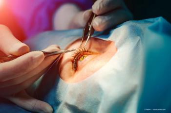
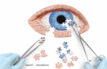
Imaging tools-such as topography, tomography, raytracing, and anterior-segment optical coherence tomography (ASOCT)-are helpful to both plan refractive surgeries and assess any potential complications, says Sonia H. Yoo, MD.
Reviewed by Sonia H. Yoo, MD
Imaging tools-such as topography, tomography, raytracing, and anterior-segment optical coherence tomography (ASOCT)-are helpful to both plan refractive surgeries and assess any potential complications, said Sonia H. Yoo, MD.
Overview of tools
Topography can be helpful for keratoconus screening, surgical planning, and astigmatism correction. It also can be used to document the effects of surgery, investigate any poor outcomes, and plan for enhancements, added Dr. Yoo, professor, Bascom Palmer Eye Institute, University of Miami Miller School of Medicine.
“It’s important to recognize pathological patterns to rule out candidates such as the patient with inferior steepening,” Dr. Yoo said.
Keratorefractive surgery performed in such cases is associated with a higher risk of keratectasia and postoperative topographic instability. A decentered myopic ablation and a central island are other complications that can be seen by topography and are associated with poor outcomes.
Tomography generates a 3-D recreation and can measure the anterior and posterior surfaces, which can be helpful with assessment of the cornea. Wavefront maps and ray-tracing help surgeons to evaluate higherorder aberrations (HOAs), which can be responsible for reduced quality of vision, Dr. Yoo said.
Ray-tracing is another tool to evaluate HOAs. Anterior-segment OCT is a noncontact approach to evaluate the cornea and anterior segment, and it can penetrate through corneal opacities and scars. One way to use AS-OCT is to see if a patient is a candidate for a LASIK enhancement.
A closer look
Dr. Yoo presented some example patients evaluated with these imaging tools. For instance, a 30-year-old male had LASIK and persistently blurry vision. By looking at topography and HOAs, Dr. Yoo could see significant amount of coma, consistent with his complaint of blurriness. Another patient was a 28-year-old male wanting to have refractive surgery.
Dr. Yoo also had treated the patient’s father, who had bilateral corneal transplants for keratoconus. The son had a spherical refraction, good best-corrected visual acuity (BCVA), and relatively normal tomography. Via wavefront, a high degree of coma was seen.
“Coma is an early finding with keratoconus and because of his strong family history, we decided to do surface ablation,” Dr. Yoo said. The patient has been happy with his result at 1-month post-op and is 20/20 J1+ in both eyes.
However, he does notice some glare at night. Dr. Yoo also shared the story of a female with a LASIK evaluation who had good BCVA and not much astigmatism but who also had a significant paracentral scar. Her topography was regular, but it also showed a thick cornea. Because of the ulcer and good BCVA, Dr. Yoo decided to do LASIK in one eye and transepithelial PRK in the eye with the scar.
“We removed 50 μm with the laser, then we did the full treatment,” Dr. Yoo said. “We informed her there could be disturbances.” However, the patient ultimately had a good outcome.
Disclosures:
Sonia H. Yoo, MD
E: [email protected]
This article was adapted from Dr. Yoo’s presentation at the 2018 meeting of the American Academy of Ophthalmology. Dr. Yoo is an equity partner in Resolve Ophthalmics and a consultant for Avedro.
Advertisement
Related Content
Advertisement
Latest CME


Advertisement
Advertisement