DMEK outcomes favorable after mid-term duration of follow-up
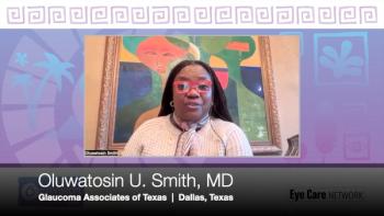
Findings from a mid-term evaluation of the first Descemet Membrane Endothelial Keratoplasty (DMEK) cohort show that the procedure results in a near complete visual recovery that is achieved at 6 months and seems to remain stable for at least up to 6 years, said Fook Chang Lam, MD.
Chicago-Findings from a mid-term evaluation of the first Descemet Membrane Endothelial Keratoplasty (DMEK) cohort show that the procedure results in a near complete visual recovery that is achieved at 6 months and seems to remain stable for at least up to 6 years, said Fook Chang Lam, MD.
The world’s first DMEK was performed by Gerrit Melles, MD, PhD, and colleagues at the Institute for Innovative Ocular Surgery, Rotterdam, The Netherlands, more than 6 years ago.
Now, data from the first 300 operated eyes followed for at least 2 years prove that the procedure continues to be effective in the mid-term, Dr. Lam said.
“Excluding eyes with low visual potential, best-corrected visual acuity was 20/40 or better in more than 95% of eyes by 6 months,” he said. “And, what is really remarkable, the number of patients achieving 20/20 and 20/17 continues to increase with the passage of time.”
He also reported safety outcomes. Endothelial cell density decreased 34% in the first 6 months after surgery. Thereafter, it declined at a steady annual rate of 8%.
The review of postoperative complications showed such events were very rare after the first 6 months of surgery, but also demonstrated the effect of the learning curve. Although the overall graft survival rate was 86%, Dr. Lam pointed out that the success rate was much higher when considering only the last 100 to 200 cases.
“Early primary graft failure occurred in four eyes, for a rate of 1.3% rate,” he said. “However, excluding three cases that occurred in our learning curve, the rate decreases to 0.04%.”
The rate of secondary glaucoma was 1.3%, and iatrogenic cataract occurred in 9.2% of phakic eyes. There was a 0.7% rate of late-onset graft failure, and graft rejection occurred at a rate of 2% from 2 to 6 years, although two-thirds of those patients regained corneal clarity with topical treatment.
Newsletter
Don’t miss out—get Ophthalmology Times updates on the latest clinical advancements and expert interviews, straight to your inbox.


